


Help ?

IGMIN: We're glad you're here. Please click 'create a new query' if you are a new visitor to our website and need further information from us.

If you are already a member of our network and need to keep track of any developments regarding a question you have already submitted, click 'take me to my Query.'

Search

Organised by  IgMin Fevicon

Regional sites

Browse by Subjects

Welcome to IgMin Research – an Open Access journal uniting Biology, Medicine, and Engineering. We’re dedicated to advancing global knowledge and fostering collaboration across scientific fields.

Browse by Sections

At IgMin Research, we bridge the frontiers of Biology, Medicine, and Engineering to foster interdisciplinary innovation. Our expanded scope now embraces a wide spectrum of scientific disciplines, empowering global researchers to explore, contribute, and collaborate through open access.

Special Issues

We foster discussions that bridge multiple fields for a more comprehensive understanding.

Members

We foster discussions that bridge multiple fields for a more comprehensive understanding.

Articles

We foster discussions that bridge multiple fields for a more comprehensive understanding.

Explore Content

We foster discussions that bridge multiple fields for a more comprehensive understanding.

Identify Us

We foster discussions that bridge multiple fields for a more comprehensive understanding.

IgMin Corporation

Welcome to IgMin, a leading platform dedicated to enhancing knowledge dissemination and professional growth across multiple fields of science, technology, and the humanities. We believe in the power of open access, collaboration, and innovation. Our goal is to provide individuals and organizations with the tools they need to succeed in the global knowledge economy.

Publications Support
[email protected]
E-Books Support
[email protected]
Webinars & Conferences Support
[email protected]
Content Writing Support
[email protected]
IT Support
[email protected]

Search

Select Language

Explore Section

Content for the explore section slider goes here.

Abstract

Abstract at IgMin Research

We foster discussions that bridge multiple fields for a more comprehensive understanding.

Engineering Group Research Article Article ID: igmin357

Computed Tomography Kidney Disease Recognition using Deep Learning

Artificial Intelligence DOI10.61927/igmin357 Affiliation

Affiliation

    Department of CSE (AI & ML), ATME College of Engineering, Mysore, India

73
VIEWS
22
DOWNLOADS
Connect with Us

Abstract

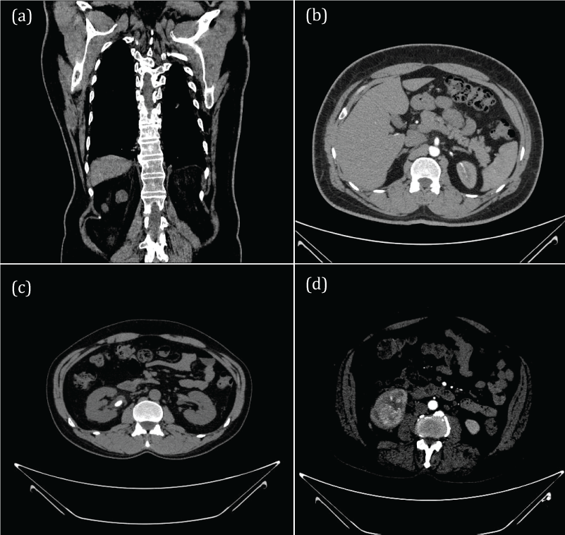
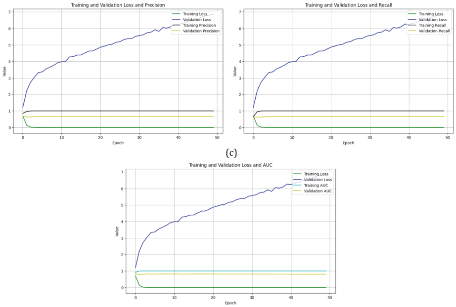
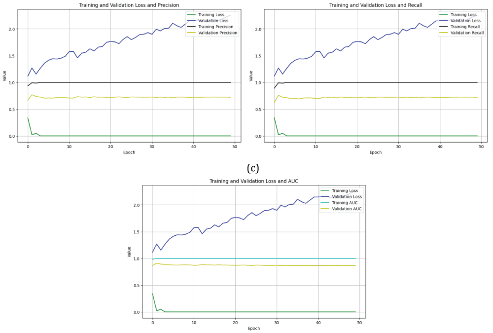
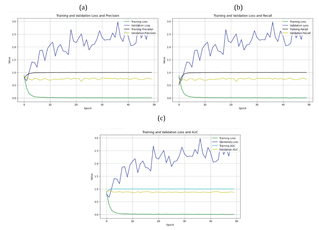
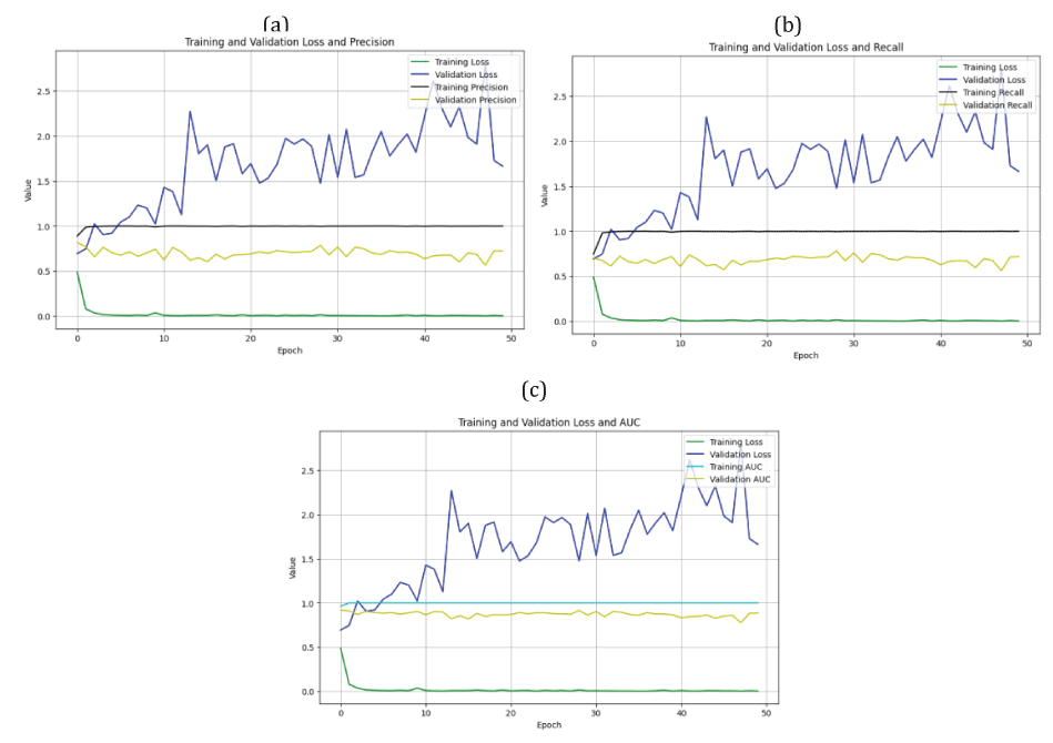
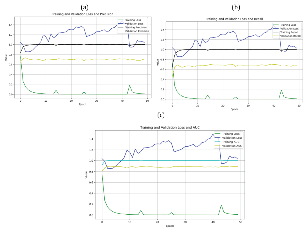
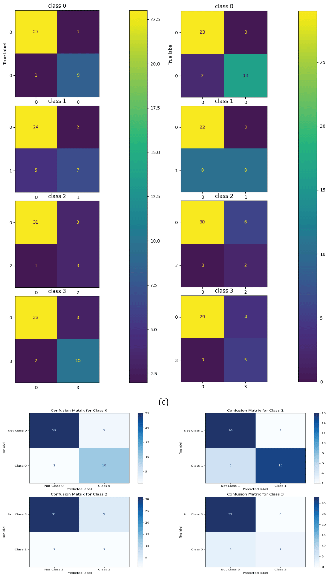
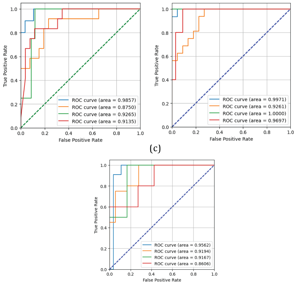
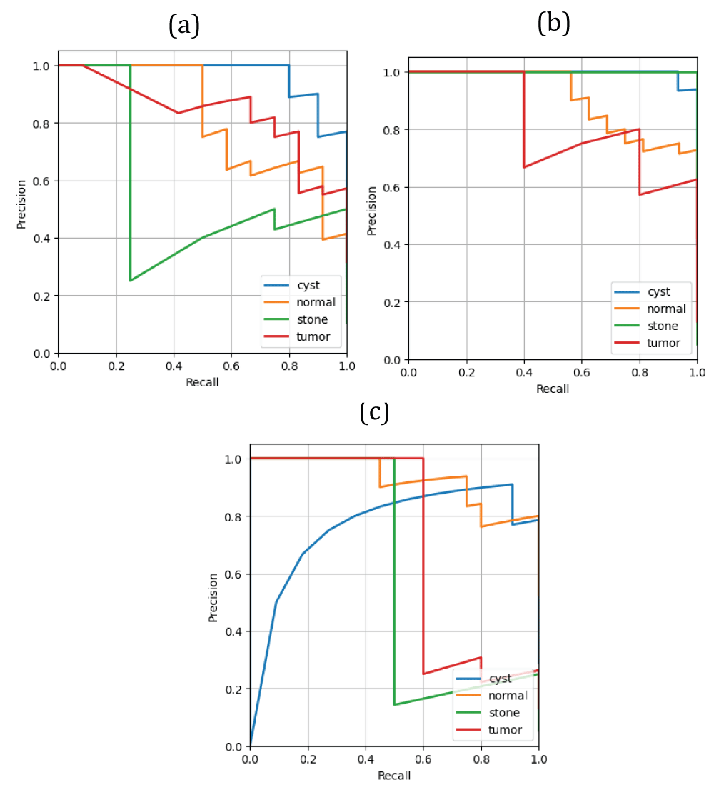
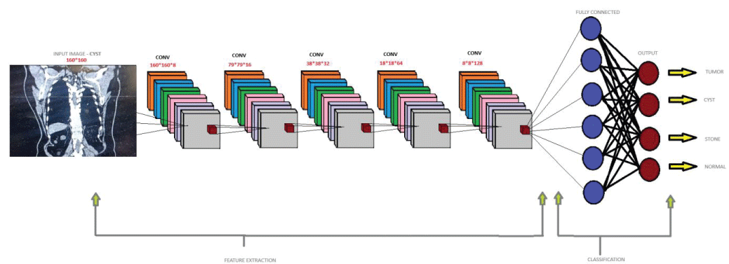
The present paper demonstrates kidney disease recognition in computed tomography (CT) images using deep neural networks, including convolutional neural networks (CNNs), the visual geometry group (VGG) network, and the deep residual network (ResNet-50). The kidney disease dataset consists of images among four categories, i.e cyst, normal, stone, and tumor. The class “cyst” consists of 3,709 images, the class “normal” consists of 5,077 images, the class “stone” consists of 1,377 images, and finally the class “tumor” consists of 2,283 images. In total, the dataset has 12,446 images. This dataset was passed through the deep CNN, VGG19, and ResNet-50 models to obtain the results. The major novelty of this work compared to previous works is that the algorithms of the CNN and ResNet-50 models have been modified, compared to existing works. The results, such as error/positive-predictive-value (PPV), error/probability of detection (POD), and error/area under the curve (AUC) curves, are shown for the confirmation of the work. Further, the error matrix, specificity/POD, and PPV/POD curves are also shown for the justification of the work. Finally, the performance metrics such as accuracy, PPV, POD, and F1-score values obtained from the error matrix are also shown for the justification of the work. The CNN model has achieved an accuracy of 0.95 for class “cyst”, 0.82 for class “normal”, 0.89 for class “stone”, and 0.87 for class “tumor”. The VGG19 neural network model has achieved an accuracy of 0.95 for class “cyst”, 0.79 for class “normal”, 0.84 for class “stone”, and 0.89 for class “tumor”. The ResNet-50 neural network model has achieved an accuracy of 0.92 for class “cyst”, 0.82 for class “normal”, 0.84 for class “stone”, and 0.92 for class “tumor”. The deep CNN model has achieved an area under the curve (AUC) value of 0.98 for class “cyst”, 0.87 for class “normal”, 0.92 for class “stone”, and 0.91 for class “tumor”. The deep CNN model has the highest AUC value for class “cyst” compared to other classes. The VGG19 model has an AUC value of 0.99 for class “cyst”, 0.92 for class “normal”, 1.00 for class “stone”, and 0.96 for class “tumor”. The VGG19 model has the highest AUC value for class “stone” compared to other classes. The ResNet-50 model has AUC values of 0.96 for class “cyst”, 0.92 for class “normal”, 0.92 for class “stone”, and 0.86 for class “tumor”. The ResNet-50 model has the highest AUC value for class “cyst” compared to the other classes.

Figures

References

    1. Farooq MS, Tariq A. A deep learning architectures for kidney disease classification. arXiv. 2024. doi:10.48550/arXiv.2403.15895
    2. Mohsen S, Ali AM, El-Rabaie ESM, ElKaseer A, Scholz SG, Hassan AMA. Brain tumor classification using hybrid single image super-resolution technique with ResNeXt101_32×8d and VGG19 pre-trained models. IEEE Access. 2023;11:55582–55595. doi:10.1109/ACCESS.2023.3281529
    3. Hossain MI, Maruf MH, Khan MAR, Prity FS, Fatema S, Ejaz MS, Khan MAS. Heart disease prediction using distinct artificial intelligence techniques: Performance analysis and comparison. Iran J Comput Sci. 2023;6(4):397–417. doi:10.1007/s42044-023-00148-7
    4. Jacobson SH. Chronic kidney disease—A public health problem? Lakartidningen. 2013;110(21):1018–1020.
    5. Jha V, Garcia-Garcia G, Iseki K, Li Z, Naicker S, Plattner B, Remuzzi G, Yang CW. Chronic kidney disease: Global dimension and perspectives. Lancet. 2013;382(9888):260–272. doi:10.1016/S0140-6736(13)60687-X
    6. Foreman KJ, Marquez N, Dolgert A, Fukutaki K, Fullman N, McGaughey M, Pletcher MA, Smith AE, Tang K, Yuan CW, Brown JC, Friedman J, He J, Heuton KR, Holmberg M, Patel DJ, Reidy P, Carter A, Cercy K, Murray CJL. Forecasting life expectancy, years of life lost, and all-cause and cause-specific mortality for 250 causes of death: Reference and alternative scenarios for 2016–40 for 195 countries and territories. Lancet. 2018;392(10159):2052–2090. doi:10.1016/S0140-6736(18)31694-5
    7. Rediger C, Guerra LA, Keays MA, Wayne C, Reddy D, Ksara S, Leonard MP. Renal cyst evolution in childhood: A contemporary observational study. J Pediatr Urol. 2019;15(2):188.e1–188.e7. doi:10.1016/j.jpurol.2018.12.012
    8. Brownstein AJ, Bin Mahmood SU, Saeyeldin A, Velasquez Mejia C, Zafar MA, Li Y, Ziganshin BA, Rizzo JA, Elefteriades JA. Simple renal cysts and bovine aortic arch: Markers for aortic disease. Open Heart. 2019;6(1):e000862. doi:10.1136/openhrt-2018-000862
    9. Sanna E, Loukogeorgakis S, Prior T, Derwig I, Paramasivam G, Choudhry M, Lees C. Fetal abdominal cysts: Antenatal course and postnatal outcomes. J Perinat Med. 2019;47(4):418–421. doi:10.1515/jpm-2018-0319
    10. Alelign T, Petros B. Kidney stone disease: An update on current concepts. Adv Urol. 2018;2018:3068365. doi:10.1155/2018/3068365
    11. Hsieh JJ, Purdue MP, Signoretti S, Swanton C, Albiges L, Schmidinger M, Heng DYC, Larkin J, Ficarra V, Renal Cell Carcinoma Working Group. Renal cell carcinoma. Nat Rev Dis Primers. 2017;3(1):17009. doi:10.1038/nrdp.2017.9
    12. Sozio SM, Pivert KA, Caskey FJ, Levin A. The state of the global nephrology workforce: A joint ASN–ERA-EDTA–ISN investigation. Kidney Int. 2021;100(5):995–1000. doi:10.1016/j.kint.2021.08.011
    13. Wasi S, Alam SB, Rahman R, Amin MA, Kobashi S. Kidney tumor recognition from abdominal CT images using transfer learning. In: 2023 IEEE 53rd International Symposium on Multiple-Valued Logic (ISMVL). IEEE; 2023. p.54–58. doi:10.1109/ISMVL57333.2023.00021
    14. Prommakhot A, Srinonchat J. VGGNet integration for kidney tumor classification. In: 2024 12th International Electrical Engineering Congress (iEECON). IEEE; 2024. p.1–6. doi:10.1109/iEECON60677.2024.10537904
    15. Brehar RD, Mitrea DA, Nedevschi S, Moisoiu T, Elec FI, Socaciu MA. Kidney tumor segmentation and grade identification in CT images. In: 2023 IEEE 19th International Conference on Intelligent Computer Communication and Processing (ICCP). IEEE; 2023. p.293–300. doi:10.1109/ICCP60212.2023.10398718
    16. Yang G, Li G, Pan T, Kong Y, Wu J, Shu H, Zhu X. Automatic segmentation of kidney and renal tumor in CT images based on 3D fully convolutional neural network with pyramid pooling module. In: 2018 24th International Conference on Pattern Recognition (ICPR). IEEE; 2018. p.3790–3795. doi:10.1109/ICPR.2018.8545143
    17. Zhao Z, Chen H, Li J, Wang L. Boundary attention u-net for kidney and kidney tumor segmentation. In: 2022 44th Annual International Conference of the IEEE Engineering in Medicine & Biology Society (EMBC). IEEE; 2022. p.1540–1543. doi:10.1109/EMBC48229.2022.9871443
    18. Islam MN, Hasan M, Hossain MK, Alam MGR, Uddin MZ, Soylu A. Vision transformer and explainable transfer learning models for auto detection of kidney cyst, stone and tumor from CT-radiography. Sci Rep. 2022;12(1):11440. doi:10.1038/s41598-022-15634-4
    19. Alzu’bi D, Abdullah M, Hmeidi I, AlAzab R, Gharaibeh M, El-Heis M, Abualigah L. Kidney tumor detection and classification based on deep learning approaches: A new dataset in CT scans. J Healthc Eng. 2022;2022(1):3861161. doi:10.1155/2022/3861161
    20. Hossain MN, Bhuiyan E, Miah MBA, Sifat TA, Muhammad Z, Masud MFA. Detection and classification of kidney disease from CT images: An automated deep learning approach. Technologies. 2025;13(11):508. doi:10.3390/technologies13110508
    21. Jahan F, Reza AS, Morol MK, Nandi D, Hossen MJ, Rahman M. Explainable deep learning for early diagnosis of chronic kidney disease from CT images in Bangladeshi patients. Sci Rep. 2026;16(1):14819. doi:10.1038/s41598-026-42654-1
    22. Kumar Y, Brar TPS, Kaur C, Singh C. A comprehensive study of deep learning methods for kidney tumor, cyst, and stone diagnostics and detection using CT images. Arch Comput Methods Eng. 2024;31(7):4163–4188. doi:10.1007/s11831-024-10112-8
    23. Vemula N, Nandan D. Kidney Disease Detection Using Computed Tomography Scans: A Deep Learning Approach with Hybrid Activation Functions Based ResNet50. In: 2025 IEEE International Conference on Advanced Computing Technologies (ICACT). IEEE; 2025. p.701–706.
    24. He K, Zhang X, Ren S, Sun J. Deep residual learning for image recognition. In: Proceedings of the IEEE Conference on Computer Vision and Pattern Recognition. IEEE; 2016. p.770–778. doi:10.1109/CVPR.2016.90
    25. Mahesh RN U, Nelleri A. Multi-class classification and multi-output regression of three-dimensional objects using artificial intelligence applied to digital holographic information. Sensors. 2023;23(3):1095. doi:10.3390/s23031095
    26. Islam MN, Hasan M, Hossain MK, Alam MGR, Uddin MZ, Soylu A. CT kidney dataset: Normal-cyst-tumor and stone [Data set]. Kaggle. 2022. Available from: https://www.kaggle.com/datasets/nazmul0087/ct-kidney-dataset-normal-cyst-tumor-and-stone
    27. Reddy BL, Uma Mahesh RN, Nelleri A. Deep convolutional neural network for three-dimensional objects classification using off-axis digital Fresnel holography. J Mod Opt. 2022;69(13):705–717. doi:10.1080/09500340.2022.2081371
    28. Uma Mahesh RN, Lokesh Reddy B, Nelleri A. Deep learning-based multi-class 3D objects classification using digital holographic complex images. In: International Conference on Futuristic Communication and Network Technologies. Singapore: Springer Nature Singapore; 2020. p.443–448. doi:10.1007/978-981-16-4625-6_43
    29. Mahesh U, Kiran B. Three-dimensional (3-D) objects classification by means of phase-only digital holographic information using Alex Network. In: 2024 International Conference on Signal Processing, Computation, Electronics, Power and Telecommunication (IConSCEPT). IEEE; 2024. p.1–5. doi:10.1109/IConSCEPT61884.2024.10627906
    30. Mahesh U, HJ HJ, Kumar H, Umrao S, DL M. Melanocytic Nevi Classification using Transfer Learning. Igmin Res. 2025;3(7):258–267. doi:10.61927/igmin307
    31. Mahesh RU, Nagaraju S. Three-dimensional (3-D) objects classification by means of phase-only digital holographic information using deep learning. In: Data science & exploration in artificial intelligence. A.A. Balkema; 2025. p.363–371. doi:10.1201/9781003587392-53
    32. Mahesh U, Madhusudhan KV. Clustering of Three-dimensional (3-D) Objects by Means of Phase-only Digital Holographic Information using Machine Learning. Igmin Res. 2025;3(10):379–386. doi:10.61927/igmin318
    33. Uma Mahesh RN, Chandrashekar P. Deep residual network for three-dimensional (3-D) objects classification using phase-only digital holographic information. J Intell Syst. 2026;35(1):20240393. doi:10.1515/jisys-2024-0393
    34. Uma Mahesh RN, Chakrasali D, Suhas Chandra Thejasvi N, Manoj Kumar C, Bharadwaj SD. Audio Signal Classification Using Deep Learning. Igmin Res. 2026 Jun 19;4(6):208–214. doi:10.61927/igmin345. Available from: igmin.link/p345
    35. Uma Mahesh RN, Shivu M, Shivu R, Vadaga Y. Multi-class Prediction of Three-dimensional Objects by means of Phase-only digital holographic information using Deep Learning. Igmin Res. 2026 Jul 08;4(7):252–260. doi:10.61927/igmin350. Available from: igmin.link/p350

Similar Articles

Why publish with us?

  • Global Visibility – Indexed in major databases

  • Fast Peer Review – Decision within 14–21 days

  • Open Access – Maximize readership and citation

  • Multidisciplinary Scope – Biology, Medicine and Engineering

  • Editorial Board Excellence – Global experts involved

  • University Library Indexing – Via OCLC

  • Permanent Archiving – CrossRef DOI

  • APC – Affordable APCs with discounts

  • Citation – High Citation Potential

Submit Your Article

Advertisement