IGMIN: We're glad you're here. Please click 'create a new query' if you are a new visitor to our website and need further information from us.
If you are already a member of our network and need to keep track of any developments regarding a question you have already submitted, click 'take me to my Query.'
Welcome to IgMin Research – an Open Access journal uniting Biology, Medicine, and Engineering. We’re dedicated to advancing global knowledge and fostering collaboration across scientific fields.
At IgMin Research, we bridge the frontiers of Biology, Medicine, and Engineering to foster interdisciplinary innovation. Our expanded scope now embraces a wide spectrum of scientific disciplines, empowering global researchers to explore, contribute, and collaborate through open access.
Welcome to IgMin, a leading platform dedicated to enhancing knowledge dissemination and professional growth across multiple fields of science, technology, and the humanities. We believe in the power of open access, collaboration, and innovation. Our goal is to provide individuals and organizations with the tools they need to succeed in the global knowledge economy.
IgMin Publications Inc., Suite 102, West Hartford, CT - 06110, USA
Medicine GroupResearch ArticleArticle ID: igmin364
Clinical and Comorbidity Profile of Metabolic Dysfunction-Associated Steatotic Liver Disease in Tertiary Care Setting: A Retrospective Comparative Study
Background: Metabolic dysfunction-associated steatotic liver disease (MASLD) is a multisystem cardiometabolic condition. Because cardiometabolic abnormalities are components of the diagnostic framework, studies describing their prevalence in MASLD must distinguish phenotypic characterization from criterion-independent association testing.
Objective: To compare the clinical, biochemical, and routine-care liver-stiffness profiles of adults with and without MASLD in a tertiary-care setting and to describe how metabolic comorbidity burden varied across imaging-defined steatosis grades.
Methods: This retrospective comparative study included 850 adults. MASLD was operationally defined by both imaging-confirmed hepatic steatosis and at least one documented cardiometabolic risk factor. Between-group comparisons used independent-samples t tests, Mann–Whitney U tests, or chi-square tests, as appropriate. Adjusted odds ratios for comorbidities were treated as exploratory and criterion-dependent because type 2 diabetes mellitus, hypertension, and dyslipidemia contributed to MASLD ascertainment. Across steatosis grades, binary outcomes were evaluated by logistic trend models and continuous outcomes by linear regression with grade coded 1–3.
Results: Of 850 patients, 432 (50.8%) met the operational MASLD definition and 418 (49.2%) had no imaging evidence of steatosis. The MASLD group had higher body mass index, aminotransferases, gamma-glutamyl transferase, triglycerides, glycated hemoglobin, and liver stiffness, and lower high-density lipoprotein cholesterol. Type 2 diabetes mellitus and dyslipidemia were more frequent in the MASLD group; however, these comparisons are partly criterion-dependent and should not be interpreted as independent validation of association. Within the MASLD group, increasing steatosis grade was accompanied by higher metabolic comorbidity burden and worsening biochemical and liver-stiffness measurements.
Conclusion: In this tertiary-care cohort, the operational MASLD definition identified patients with an adverse hepatic and cardiometabolic profile. The graded patterns across steatosis severity support integrated metabolic and liver-risk assessment, but the retrospective cross-sectional design, incorporation of cardiometabolic factors into case definition, and non-histologic nature of liver-stiffness measurements limit causal and criterion-independent interpretation.
Metabolic dysfunction-associated steatotic liver disease (MASLD) has emerged as one of the most common chronic liver diseases worldwide and is now recognized as a major challenge in hepatology and metabolic medicine. Formerly discussed under the nomenclature of non-alcoholic fatty liver disease (NAFLD) and later metabolic dysfunction-associated fatty liver disease (MAFLD), the current MASLD framework reflects a more clinically relevant understanding of hepatic steatosis as part of systemic metabolic dysfunction rather than as an isolated liver disorder defined primarily by exclusion criteria [11Eslam M, Newsome PN, Sarin SK, Anstee QM, Targher G, Romero-Gomez M, et al. A new definition for metabolic dysfunction-associated fatty liver disease: An international expert consensus statement. J Hepatol. 2020;73(1):202–209. doi:10.1016/j.jhep.2020.03.039-44Tilg H, Effenberger M. From NAFLD to MAFLD: When pathophysiology succeeds. Nat Rev Gastroenterol Hepatol. 2020;17(7):387–388. doi:10.1038/s41575-020-0316-6]. In this context, MASLD is closely associated with obesity, insulin resistance, type 2 diabetes mellitus (T2DM), dyslipidemia, and the broader spectrum of metabolic syndrome, and it contributes substantially to both hepatic and extrahepatic morbidity [55Fouad Y, Waked I, Bollipo S, Gomaa A, Ajlouni Y, Attia D. The NAFLD-MAFLD debate: A global perspective. J Hepatol. 2021;74(6):1094–1096. doi:10.1016/j.jhep.2020.12.019-77Kaya E, Yilmaz Y. Epidemiology, natural history, and diagnosis of metabolic dysfunction-associated fatty liver disease: a comparative review with nonalcoholic fatty liver disease. Ther Adv Endocrinol Metab. 2022 Dec 10;13:20420188221139650. doi: 10.1177/20420188221139650. PMID: 36533185; PMCID: PMC9747887.].
The clinical significance of MASLD extends well beyond the risk of progressive liver injury. In addition to progression toward steatohepatitis, fibrosis, cirrhosis, and hepatocellular carcinoma, MASLD has been consistently linked to increased risks of cardiovascular disease, chronic kidney disease, and selected extrahepatic malignancies, with important implications for long-term all-cause and cardiovascular mortality [88Younossi ZM, Rinella ME, Sanyal AJ, Harrison SA, Brunt EM, Goodman Z, et al. From NAFLD to MAFLD: Implications of a premature change in terminology. Hepatology. 2021;73(3):1194–1198. doi:10.1002/hep.31420,99Eslam M, Sarin SK, Wong VW, Fan JG, Kawaguchi T, Ahn SH, et al. The Asian Pacific Association for the Study of the Liver clinical practice guidelines for the diagnosis and management of metabolic associated fatty liver disease. Hepatol Int. 2020;14(6):889–919. doi:10.1007/s12072-020-10094-2,1212Targher G, Corey KE, Byrne CD, Roden M. The complex link between NAFLD and type 2 diabetes mellitus—mechanisms and treatments. Nat Rev Gastroenterol Hepatol. 2021;18(9):599–612. doi:10.1038/s41575-021-00448-y]. This broader burden reflects the central role of metabolic multimorbidity, particularly T2DM, dyslipidemia, and hypertension, which commonly coexist with hepatic steatosis and may influence both disease severity and prognosis [1010Eslam M, Ahmed A, Després JP, Dixon JB, Foufelle F, Gastaldelli A, et al. Incorporating fatty liver disease in multidisciplinary care and novel clinical trial designs for patients with metabolic diseases. Lancet Gastroenterol Hepatol. 2021;6(9):743–753. doi:10.1016/S2468-1253(21)00132-1,1111Younossi Z, Anstee QM, Marietti M, Hardy T, Henry L, Eslam M, et al. Global burden of NAFLD and NASH: Trends, predictions, risk factors and prevention. Nat Rev Gastroenterol Hepatol. 2018;15(1):11–20. doi:10.1038/nrgastro.2017.109]. Accordingly, comprehensive comorbidity profiling and integration of hepatology care with cardiometabolic risk assessment have become increasingly important in contemporary clinical practice [66Cusi K, Isaacs S, Barb D, Basu R, Caprio S, Garvey WT, et al. A new era in the management of MAFLD. Nat Rev Gastroenterol Hepatol. 2021;18(3):196–208. doi:10.1038/s41575-020-00396-z,1313Targher G, Day CP, Bonora E. Risk of cardiovascular disease in patients with nonalcoholic fatty liver disease. N Engl J Med. 2010;363:1341–50. doi:10.1056/NEJMra0912063].
Accurate diagnosis and meaningful risk stratification are essential for the evaluation of patients with suspected MASLD. Although liver biopsy remains the histopathological reference standard for assessing steatosis, necroinflammatory activity, and fibrosis, its routine use is limited by invasiveness, sampling variability, cost, and procedure-related risk [1414Chalasani N, Younossi Z, Lavine JE, Charlton M, Cusi K, Rinella M, et al. The diagnosis and management of nonalcoholic fatty liver disease: Practice guidance from the American Association for the Study of Liver Diseases. Hepatology. 2018;67(1):328–357. doi:10.1002/hep.29367]. As a result, non-invasive approaches now play a central role in routine care. Ultrasonography remains the most widely used first-line modality for detecting hepatic steatosis because of its availability, low cost, and practicality in large clinical populations, although its sensitivity is lower in mild steatosis and it remains operator-dependent [1515Hernaez R, Lazo M, Bonekamp S, Kamel I, Brancati FL, Guallar E, Clark JM. Diagnostic accuracy and reliability of ultrasonography for the detection of fatty liver: A meta-analysis. Hepatology. 2011;54(3):1082–1090. doi:10.1002/hep.24452-1717Dulai PS, Singh S, Patel J, Soni M, Prokop LJ, Younossi Z, et al. Increased risk of mortality by fibrosis stage in nonalcoholic fatty liver disease: Systematic review and meta-analysis. Hepatology. 2017;65(5):1557–1565. doi:10.1002/hep.29085]. Current liver society guidance therefore emphasizes structured diagnostic pathways and stepwise risk stratification using non-invasive tests, including serum-based indices and elastography-based assessment, to identify patients at increased risk of clinically significant fibrosis who require specialist follow-up and more intensive management [1818Romero-Gómez M, Zelber-Sagi S, Trenell M, Cusi K, Cortez-Pinto H, Marchesini G. Treatment of MAFLD: Lifestyle and diet modification. J Hepatol. 2021;75(4):1002–1013. doi:10.1016/j.jhep.2021.07.022,1919Kaya E, Yilmaz Y. Metabolic-associated fatty liver disease (MAFLD): A multisystemic disease beyond the liver. Zhonghua Gan Zang Bing Za Zhi. 2025;33(1):77–87. doi:10.3760/cma.j.cn501113-20250103-00003. PMID: 39929687.].
Despite the growing clinical importance of MASLD, important gaps remain in the real-world characterization of affected patients, especially in tertiary-care settings where disease complexity and comorbidity burden may be higher than in community-based populations. Many published studies have focused on single risk factors, selected metabolic traits, or narrowly defined subgroups, whereas fewer have provided an integrated comparison of demographic features, biochemical abnormalities, liver stiffness, and comorbidity burden between adults with and without steatotic liver disease in routine specialist practice. This limitation is particularly relevant in the Russian Federation and similar settings, where the epidemiological burden appears to parallel global trends in obesity and metabolic syndrome, but where comprehensive tertiary-care data remain comparatively limited [2525Drapkina, O.; Ivashkin, V.. Risk factors metabolic syndrome and non-alcoholic fatty liver disease in Russian federation in national-wide DIREG-L-01903 STUDY: PP.9.238. Journal of Hypertension 29():p e217-e218, June 2011.-2727Ivashkin VT, Maev IV, Korochanskaya NV, et al. Metabolic syndrome and nonalcoholic fatty liver disease in the Russian Federation: Prevalence and clinical characteristics. Klin Med (Mosk). 2017;95(3):179–186. Russian.].
Against this background, we conducted a retrospective comparative study in a tertiary-care setting to evaluate demographic characteristics, biochemical parameters, liver stiffness, and metabolic and gastrointestinal comorbidities among adults with imaging-defined steatotic liver disease. We further examined whether increasing steatosis severity was associated with a progressively greater burden of metabolic and hepatic abnormalities. By providing a structured real-world profile in routine tertiary-care practice, this study aims to clarify whether hepatic steatosis identifies a clinically distinct high-risk metabolic phenotype and to support more risk-based, multidisciplinary approaches to patient assessment and management.
In alignment with the latest multi-society consensus guidelines, this study adopts the current MASLD nomenclature, which supersedes the previous non-alcoholic fatty liver disease (NAFLD) and metabolic dysfunction-associated fatty liver disease (MAFLD) terminologies. The transition to MASLD reflects a positive diagnostic framework, defining the condition by the presence of hepatic steatosis alongside at least one cardiometabolic risk factor, rather than relying on exclusion criteria. We utilized this updated MASLD framework to accurately capture the systemic metabolic dysregulation inherent to the disease and to ensure our tertiary-care cohort is classified according to contemporary international standards.
Against this background, we conducted a retrospective comparative study in a tertiary-care setting to compare demographic characteristics, biochemical parameters, routine-care liver-stiffness measurements, and comorbidity profiles among adults with and without imaging-defined hepatic steatosis who were classified within the contemporary MASLD framework. We also examined whether increasing imaging-defined steatosis severity was accompanied by progressively greater metabolic and hepatic abnormalities. Because cardiometabolic risk factors were required for the operational diagnosis of MASLD, analyses of type 2 diabetes mellitus, hypertension, and dyslipidemia were prespecified as descriptive and criterion-dependent rather than as independent tests of etiologic association.
This retrospective comparative study was conducted at Botkin Moscow City Clinical Hospital, a large multispecialty tertiary-care center in Moscow, Russian Federation, in collaboration with Sechenov First Moscow State Medical University. The study was designed to compare demographic characteristics, anthropometric measures, laboratory parameters, liver stiffness, and comorbidity profiles between adults with and without imaging-defined hepatic steatosis within the contemporary MASLD framework. The source dataset was derived from electronic health records of patients followed over the period from January 2020 to October 2024. After application of eligibility criteria and review of data completeness, 850 adult patients were included in the final analytical cohort, comprising 432 patients with steatotic liver disease and 418 patients without imaging evidence of steatosis.
Study population and eligibility criteria
Eligible participants were adults aged 18 years or older who had available abdominal imaging suitable for assessment of hepatic steatosis together with sufficient demographic, clinical, and biochemical data for group classification and comparative analysis. Inclusion required documentation of core study variables, including age, sex, body mass index, relevant laboratory indices, and information on major metabolic and gastrointestinal comorbidities. Patients were excluded if medical records were incomplete to the extent that reliable determination of steatosis status or key study variables was not possible. The final study population was therefore categorized into a steatosis group and a comparator group without imaging evidence of hepatic steatosis.
Disease definition and diagnostic assessment
MASLD required the presence of both (1) hepatic steatosis documented by abdominal ultrasonography and (2) at least one cardiometabolic risk factor documented in the medical record. The available qualifying risk factors were increased body mass index or obesity, type 2 diabetes mellitus or abnormal glycemia, hypertension, and dyslipidemia, as defined from diagnoses, medications, and available clinical or laboratory thresholds. The comparator group had no imaging evidence of hepatic steatosis. Because type 2 diabetes mellitus, hypertension, and dyslipidemia contributed to MASLD case ascertainment, subsequent between-group analyses of these conditions are not independent of the operational disease definition and were therefore interpreted as descriptive, criterion-linked comparisons.
Hepatic steatosis was assessed by abdominal ultrasonography performed during routine clinical care. Steatosis was identified from standard sonographic features, including increased hepatic echogenicity relative to the renal cortex, vascular blurring, and posterior beam attenuation. For severity analyses, radiology reports describing mild, moderate, or severe steatosis were recorded as Grades 1, 2, and 3, respectively; descriptive reports were harmonized to these ordinal categories using a predefined rubric.
For severity-based analyses, steatosis grade was assigned from radiology reports. Reports explicitly describing steatosis as mild, moderate, or severe were recorded directly as Grades 1, 2, and 3, respectively. When descriptive rather than formal grading terminology was used, report language was harmonized into ordinal severity categories according to a predefined rubric based on conventional ultrasonographic descriptors. Liver stiffness was analyzed as a non-invasive biomechanical measurement and was not considered equivalent to histologic fibrosis stage. Values may be influenced by inflammation, cholestasis, venous congestion, food intake, and technical factors; therefore, they were interpreted as supportive risk-stratification data rather than direct histologic confirmation of fibrosis.
Data collection and study variables
Clinical and laboratory data were extracted from electronic health records using a predefined data collection framework. Recorded variables included age, sex, body mass index, alanine aminotransferase, aspartate aminotransferase, gamma-glutamyl transferase, glycated hemoglobin, triglycerides, high-density lipoprotein cholesterol, liver stiffness, and documented comorbid conditions. Demographic, anthropometric, biochemical, elastographic, and comorbidity data were reviewed for completeness before inclusion in analysis.
Comorbidities were identified through integration of ICD-10 diagnostic codes, clinician-documented diagnoses, medication history, and relevant clinical or laboratory thresholds available in the medical record. Hypertension was defined by a documented diagnosis and/or blood pressure measurements consistent with hypertension in routine clinical practice. Type 2 diabetes mellitus was defined by a documented diagnosis, use of antidiabetic therapy, fasting plasma glucose of at least 7.0 mmol/L, or glycated hemoglobin of at least 6.5%. Dyslipidemia was defined by a documented diagnosis, use of lipid-lowering therapy, or lipid abnormalities including total cholesterol of at least 5.2 mmol/L, low-density lipoprotein cholesterol of at least 3.4 mmol/L, or triglycerides of at least 1.7 mmol/L. Gastroesophageal reflux disease, chronic pancreatitis, and other recorded comorbidities were identified from clinician documentation and diagnostic coding in routine care. Where feasible, diagnoses were cross-checked across more than one data source to improve classification reliability and to reduce duplicate capture of overlapping diagnostic labels.
Statistical analysis
Continuous variables were summarized as mean ± standard deviation when approximately normally distributed and as median with interquartile range when distributional assumptions were not met. Categorical variables were reported as number (percentage). Between-group comparisons used independent-samples t tests or Mann–Whitney U tests for continuous variables and Pearson chi-square tests for categorical variables, as appropriate.
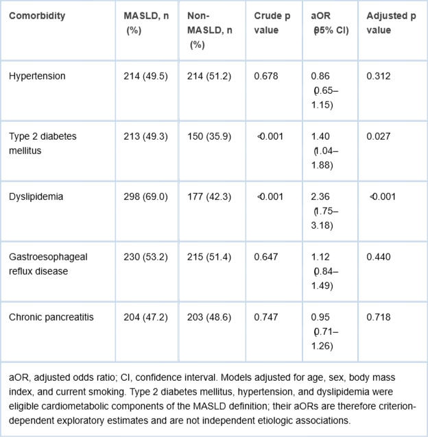
Exploratory multivariable logistic regression models were used to describe covariate-adjusted co-occurrence of MASLD status and recorded comorbidities after adjustment for age, sex, body mass index, and current smoking. Adjusted odds ratios (aORs) are reported with 95% confidence intervals. Because type 2 diabetes mellitus, hypertension, and dyslipidemia were eligible cardiometabolic components of the MASLD definition, estimates for these variables are criterion-dependent and were not interpreted as independent evidence of association or causation.
For ordinal analyses across steatosis Grades 1–3, grade was entered as a continuous variable coded 1, 2, and 3. Binary comorbidities were evaluated using univariable binomial logistic regression; results are reported as the odds ratio per one-grade increase, 95% confidence interval, Wald z statistic, and p value. Continuous outcomes were evaluated using ordinary least-squares linear regression; results are reported as the regression coefficient per one-grade increase (β), 95% confidence interval, t statistic with degrees of freedom, and p value. These grade-based comorbidity analyses describe within-cohort co-distribution and do not eliminate the criterion-dependence inherent in MASLD classification.
All tests were two-sided, and p < 0.05 was considered statistically significant. Exact p values are reported to three decimal places when p ≥ 0.001; smaller values are reported as p < 0.001. Analyses were performed primarily in SPSS version 26.0, with selected analytical checks reproduced in Python.
Ethical considerations
The study was based on a retrospective analysis of routinely collected hospital data. It was conducted in accordance with the Declaration of Helsinki and approved by the Local Ethics Committee of Sechenov First Moscow State Medical University in collaboration with the Botkin Moscow City Clinical Hospital team (Extract from Protocol No. 27-24; approval date 07 November 2024). Ethical consent and approval were granted for the entire period up to the completion of the study. Because this was a strictly retrospective study extracting historical data from electronic medical databases for patients followed over the given period, there was no direct, in-person contact with any patients. All data were derived directly from the electronic database and de-identified prior to analysis; therefore, the requirement for informed patient consent was formally waived by the ethics committee.
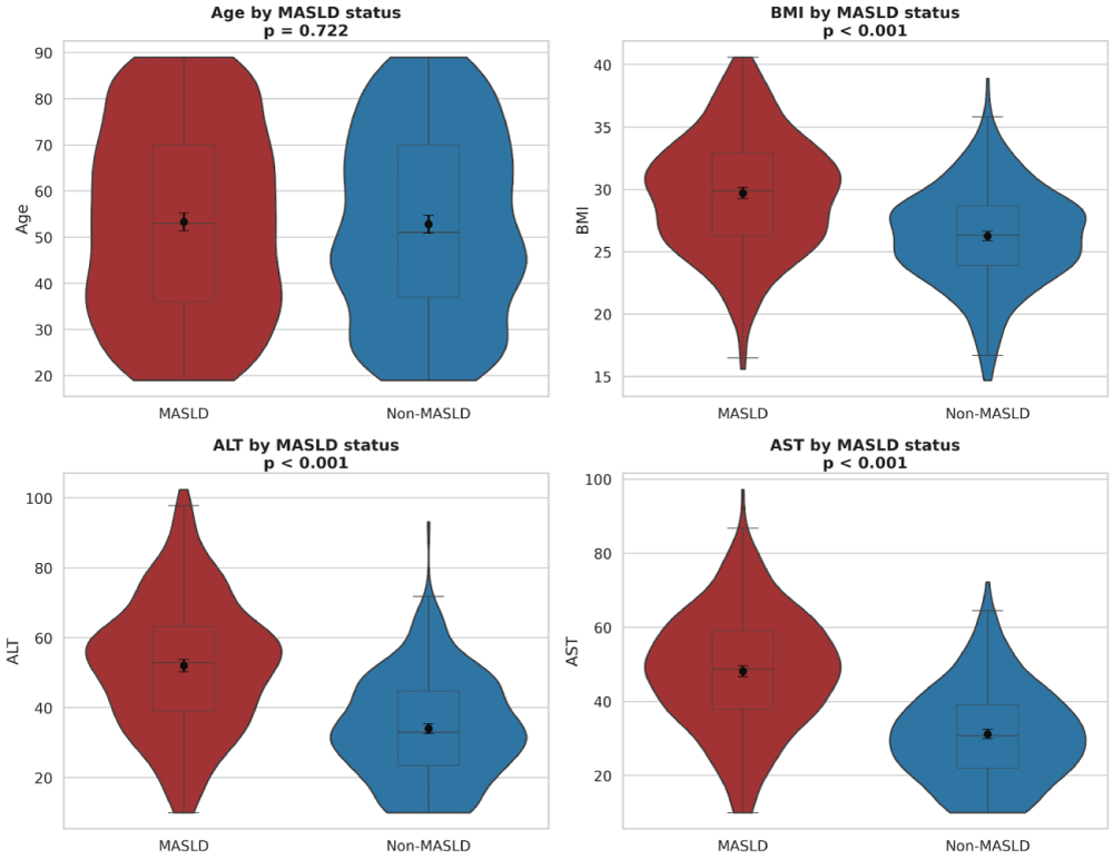
A total of 850 patients were included in the analysis, comprising 432 patients with MASLD and 418 without MASLD. Baseline demographic, lifestyle, biochemical, and elastographic characteristics are presented in Table 1. The cohorts were successfully matched by gender, with males comprising 48.6% of the MASLD group and 49.5% of the non-MASLD group (p = 0.790). Mean age was also similar between groups (53.31 ± 20.53 years in the MASLD group vs. 52.81 ± 20.02 years in the non-MASLD group, p = 0.722).
Table 1: Baseline demographic, anthropometric, biochemical, metabolic, and elastographic characteristics of the study population stratified by MASLD status.
Figure 1: Distribution of demographic and key biochemical parameters by MASLD status. Violin plots overlaid with box plots compare age, body mass index, ALT, and AST between patients with MASLD and those without MASLD. The width of each violin reflects data density, boxes indicate the interquartile range, center lines indicate medians, and overlaid markers denote group means with 95% confidence intervals. Age distributions largely overlap, whereas body mass index and liver enzyme distributions are shifted upward in MASLD, indicating greater adiposity and biochemical evidence of hepatic injury.
Regarding anthropometric and lifestyle factors, patients with MASLD had a substantially higher body mass index than those without MASLD (29.71 ± 4.62 vs. 26.25 ± 3.90 kg/m², p < 0.001), which translated to a markedly higher prevalence of clinical obesity (49.3% vs. 15.8%, p < 0.001). Furthermore, current smoking was significantly more prevalent in the MASLD cohort (32.6% vs. 24.9%, p = 0.013), and reported alcohol intake patterns differed significantly between the groups (p < 0.001).
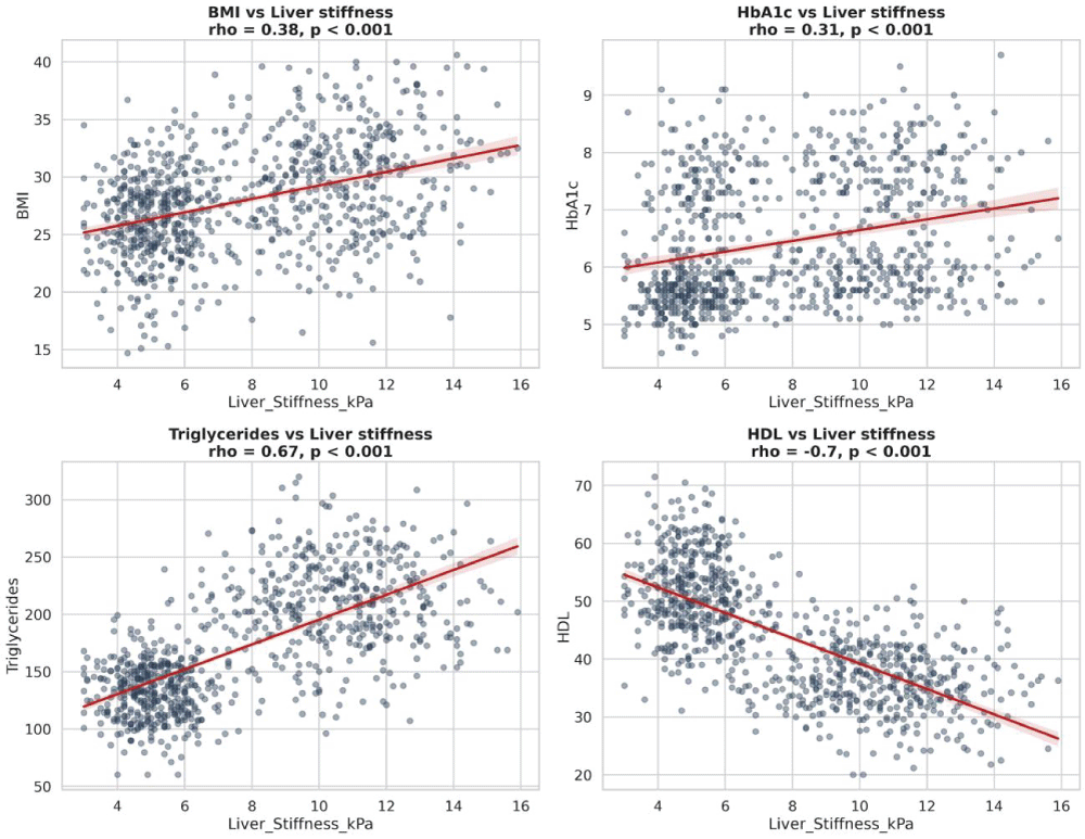
Markers of hepatic injury were higher in the MASLD group, including alanine aminotransferase (52.01 ± 18.53 vs. 34.05 ± 14.56 U/L), aspartate aminotransferase (48.12 ± 15.64 vs. 31.21 ± 12.70 U/L), and gamma-glutamyl transferase (62.19 ± 11.92 vs. 29.43 ± 7.81 U/L; all p < 0.001). The MASLD group also had higher glycated hemoglobin and triglycerides and lower high-density lipoprotein cholesterol (all p < 0.001). Routine-care liver-stiffness measurements were higher in the MASLD group (10.54 ± 2.00 vs. 5.08 ± 0.94 kPa, p < 0.001); this difference reflects a non-invasive stiffness measure and should not be interpreted as direct equivalence to histologic fibrosis stage.
Comorbidity associations
The distribution of recorded comorbidities is summarized in revised Table 2. Type 2 diabetes mellitus was documented in 49.3% of patients with MASLD and 35.9% of patients without imaging-defined steatosis; the exploratory adjusted estimate was aOR 1.40 (95% CI 1.04 - 1.88; p = 0.027). Dyslipidemia was documented in 69.0% and 42.3%, respectively, with an exploratory aOR of 2.36 (95% CI 1.75 - 3.18; p < 0.001). These estimates quantify criterion-linked co-occurrence and must not be interpreted as independent confirmation that MASLD predicts either condition, because both conditions could contribute to MASLD classification.
Table 2: Comorbidity prevalence and exploratory adjusted associations by MASLD status.
Hypertension was documented in 49.5% of patients with MASLD and 51.2% of patients without steatosis (aOR 0.86, 95% CI 0.65–1.15; p = 0.312). Gastroesophageal reflux disease and chronic pancreatitis also did not differ after adjustment. Unlike the cardiometabolic criteria, these gastrointestinal conditions were not components of the MASLD definition; nevertheless, the cross-sectional design precludes causal interpretation.
Figure 2: Relationships between routine-care liver-stiffness measurements and metabolic parameters. Scatter plots with fitted regression lines and 95% confidence bands show the cross-sectional relationships of liver stiffness with body mass index, glycated hemoglobin, triglycerides, and high-density lipoprotein cholesterol. These unadjusted visual associations are descriptive, do not establish causality, and should not be interpreted as direct measures of histologic fibrosis.
Relationship between steatosis grade and metabolic comorbidities
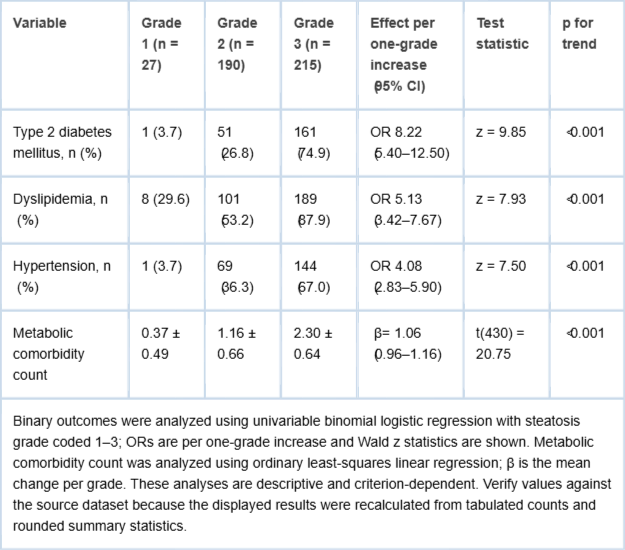
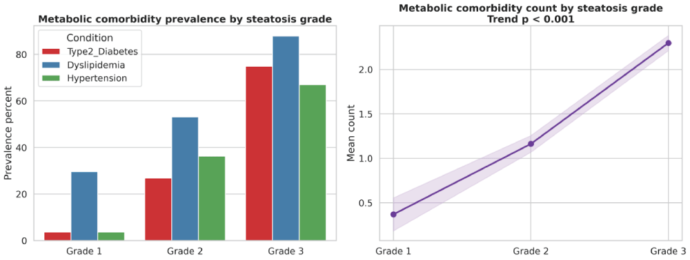
Among patients with MASLD, steatosis severity demonstrated a strong dose-response relationship with metabolic comorbidity burden (Table 3). The prevalence of type 2 diabetes increased from 3.7% in Grade 1 steatosis to 26.8% in Grade 2 and 74.9% in Grade 3. Dyslipidemia increased in parallel from 29.6% to 53.2% and 87.9% across Grades 1, 2, and 3, respectively. Hypertension also rose markedly across the same grades, from 3.7% in Grade 1 to 36.3% in Grade 2 and 67.0% in Grade 3. Consistent with these individual comorbidities, the mean metabolic comorbidity count increased progressively from 0.37 ± 0.49 in Grade 1 to 1.16 ± 0.66 in Grade 2 and 2.30 ± 0.64 in Grade 3, indicating a highly significant monotonic trend. These findings demonstrate a clear graded relationship between worsening steatosis and increasing metabolic multimorbidity.
Table 3: Metabolic comorbidity burden across imaging-defined steatosis grades among patients with MASLD.
Figure 3: Metabolic comorbidity burden across imaging-defined steatosis grades among patients with MASLD. Bars show the prevalence of type 2 diabetes mellitus, dyslipidemia, and hypertension, and the line shows the mean number of metabolic comorbidities. The ordered pattern describes within-cohort co-distribution; because these conditions could contribute to MASLD classification, the figure does not demonstrate a criterion-independent or causal relationship.
Within the MASLD group, increasing imaging-defined steatosis grade was accompanied by a higher prevalence of type 2 diabetes mellitus, dyslipidemia, and hypertension (revised Table 3). In logistic trend models, the odds per one-grade increase were 8.22 for type 2 diabetes mellitus (95% CI 5.40–12.50; z = 9.85; p < 0.001), 5.13 for dyslipidemia (95% CI 3.42–7.67; z = 7.93; p < 0.001), and 4.08 for hypertension (95% CI 2.83–5.90; z = 7.50; p < 0.001). The mean metabolic comorbidity count increased by 1.06 conditions per grade (95% CI 0.96–1.16; t(430) = 20.75; p < 0.001). These results show a strong monotonic co-distribution within the MASLD cohort, but they remain criterion-dependent and do not establish a causal effect of steatosis severity on cardiometabolic disease.
Table 3. Prevalence of major metabolic comorbidities according to imaging-defined steatosis grade among patients with MASLD. Data are presented as percentages for individual comorbidities and mean ± standard deviation for the total metabolic comorbidity count. This table demonstrates the progressive accumulation of metabolic disease burden with increasing steatosis severity.
Binary outcomes were analyzed using univariable binomial logistic regression with steatosis grade coded 1–3; ORs are per one-grade increase and Wald z statistics are shown. Metabolic comorbidity count was analyzed using ordinary least-squares linear regression; β is the mean change per grade. These analyses are descriptive and criterion-dependent. Verify values against the source dataset because the displayed results were recalculated from tabulated counts and rounded summary statistics.
Biochemical and liver stiffness parameters by steatosis grade
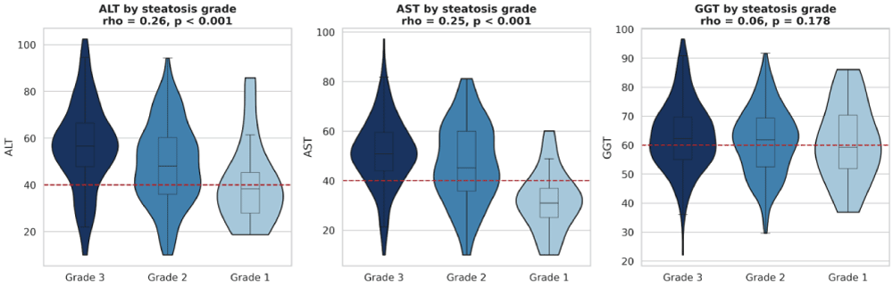
Biochemical and liver-stiffness measurements also varied across steatosis grades (revised Table 4). Per one-grade increase, alanine aminotransferase increased by 7.63 U/L (95% CI 4.84–10.42; t(430) = 5.38; p < 0.001), aspartate aminotransferase by 6.97 U/L (95% CI 4.63–9.31; t(430) = 5.85; p < 0.001), glycated hemoglobin by 0.75 percentage points (95% CI 0.60–0.90; t(430) = 9.77; p < 0.001), and triglycerides by 10.97 mg/dL (95% CI 4.94–16.99; t(430) = 3.58; p < 0.001). High-density lipoprotein cholesterol decreased by 1.46 mg/dL per grade (95% CI −2.37 to −0.55; t(430) = −3.16; p = 0.002). Liver stiffness increased by 0.82 kPa per grade (95% CI 0.52–1.12; t(430) = 5.36; p < 0.001). Gamma-glutamyl transferase did not show a statistically significant linear grade trend (β = 1.41 U/L per grade, 95% CI −0.44 to 3.26; t(430) = 1.50; p = 0.134).
Table 4: Biochemical, metabolic, and liver-stiffness parameters across imaging-defined steatosis grades among patients with MASLD.
Figure 4: Distribution of liver biochemical markers across imaging-defined steatosis grades. Violin plots with overlaid box plots compare alanine aminotransferase, aspartate aminotransferase, and gamma-glutamyl transferase across Grades 1–3. Horizontal reference lines indicate approximate upper limits of normal. Alanine and aspartate aminotransferase show ordered upward shifts, whereas gamma-glutamyl transferase does not show a statistically significant linear trend (p = 0.134).
Overall, the grade-based analyses demonstrate ordered cross-sectional patterns in metabolic, biochemical, and non-invasive liver-stiffness measurements. They should not be interpreted as evidence of temporal progression, causation, or histologically confirmed fibrosis.
Taken together, these analyses show that MASLD in our study population is characterized by a markedly worse metabolic and hepatic profile than the absence of MASLD. In addition, steatosis severity is closely linked to the burden of metabolic comorbidities and to worsening biochemical and elastographic parameters, supporting a clinically meaningful dose-response relationship within MASLD severity strata.
In this single-center tertiary-care cohort, adults meeting the operational MASLD definition had greater adiposity, higher aminotransferases, glycated hemoglobin, triglycerides, and routine-care liver-stiffness measurements, and lower high-density lipoprotein cholesterol than adults without imaging evidence of steatosis. Increasing steatosis grade was accompanied by ordered increases in metabolic comorbidity burden and several biochemical and liver-stiffness measurements. These findings characterize the clinical phenotype observed in this referral population but do not establish temporal or causal relationships [11Eslam M, Newsome PN, Sarin SK, Anstee QM, Targher G, Romero-Gomez M, et al. A new definition for metabolic dysfunction-associated fatty liver disease: An international expert consensus statement. J Hepatol. 2020;73(1):202–209. doi:10.1016/j.jhep.2020.03.039,33Eslam M, Sanyal AJ, George J. MAFLD: A consensus-driven proposed nomenclature for metabolic associated fatty liver disease. Gastroenterology. 2020;158(7):1999–2014. doi:10.1053/j.gastro.2019.11.312,44Tilg H, Effenberger M. From NAFLD to MAFLD: When pathophysiology succeeds. Nat Rev Gastroenterol Hepatol. 2020;17(7):387–388. doi:10.1038/s41575-020-0316-6,66Cusi K, Isaacs S, Barb D, Basu R, Caprio S, Garvey WT, et al. A new era in the management of MAFLD. Nat Rev Gastroenterol Hepatol. 2021;18(3):196–208. doi:10.1038/s41575-020-00396-z].
A central interpretive issue is incorporation bias. MASLD was defined by imaging-confirmed steatosis together with at least one cardiometabolic risk factor; therefore, comparisons of type 2 diabetes mellitus, hypertension, and dyslipidemia between MASLD and non-steatosis groups are partly determined by the case definition itself [55Fouad Y, Waked I, Bollipo S, Gomaa A, Ajlouni Y, Attia D. The NAFLD-MAFLD debate: A global perspective. J Hepatol. 2021;74(6):1094–1096. doi:10.1016/j.jhep.2020.12.019,88Younossi ZM, Rinella ME, Sanyal AJ, Harrison SA, Brunt EM, Goodman Z, et al. From NAFLD to MAFLD: Implications of a premature change in terminology. Hepatology. 2021;73(3):1194–1198. doi:10.1002/hep.31420,99Eslam M, Sarin SK, Wong VW, Fan JG, Kawaguchi T, Ahn SH, et al. The Asian Pacific Association for the Study of the Liver clinical practice guidelines for the diagnosis and management of metabolic associated fatty liver disease. Hepatol Int. 2020;14(6):889–919. doi:10.1007/s12072-020-10094-2]. The adjusted odds ratios for these variables should consequently be understood as descriptive estimates of criterion-linked co-occurrence rather than independent evidence that MASLD causes, predicts, or is independently associated with the defining cardiometabolic conditions [4-64-6Tilg H, Effenberger M. From NAFLD to MAFLD: When pathophysiology succeeds. Nat Rev Gastroenterol Hepatol. 2020;17(7):387–388. doi:10.1038/s41575-020-0316-6,1010Eslam M, Ahmed A, Després JP, Dixon JB, Foufelle F, Gastaldelli A, et al. Incorporating fatty liver disease in multidisciplinary care and novel clinical trial designs for patients with metabolic diseases. Lancet Gastroenterol Hepatol. 2021;6(9):743–753. doi:10.1016/S2468-1253(21)00132-1].
The observed prevalence of type 2 diabetes mellitus and dyslipidemia remains clinically informative as a description of the tertiary-care MASLD phenotype. However, biological plausibility and consistency with prior literature do not remove the definitional circularity in the present cross-sectional analysis. Accordingly, the study supports systematic assessment of glycemic and lipid abnormalities among patients with steatotic liver disease, while criterion-independent associations should be evaluated in future studies using alternative case definitions, sensitivity analyses that exclude the outcome under study from the qualifying criterion, or longitudinal designs.
The grade-based findings provide an additional descriptive signal. Higher steatosis grades were accompanied by progressively higher metabolic comorbidity counts, aminotransferases, glycated hemoglobin, triglycerides, and liver-stiffness values, together with lower high-density lipoprotein cholesterol. Because the analysis was cross-sectional, the monotonic pattern does not demonstrate disease progression over time. In addition, liver stiffness is influenced by technical and clinical factors and cannot be equated directly with histologic fibrosis. It is therefore most appropriately interpreted here as a non-invasive risk-stratification measure.
Clinically, the findings support integrated evaluation of liver-related and cardiometabolic risk in patients with MASLD. The data favor severity-oriented assessment rather than reliance on binary steatosis status alone, but management implications should be based on validated non-invasive pathways, complete metabolic evaluation, and clinical context rather than on the present observational associations in isolation.
These findings support the current view that MASLD should be understood as a multisystem cardiometabolic disorder rather than a purely hepatic disease entity [44Tilg H, Effenberger M. From NAFLD to MAFLD: When pathophysiology succeeds. Nat Rev Gastroenterol Hepatol. 2020;17(7):387–388. doi:10.1038/s41575-020-0316-6,66Cusi K, Isaacs S, Barb D, Basu R, Caprio S, Garvey WT, et al. A new era in the management of MAFLD. Nat Rev Gastroenterol Hepatol. 2021;18(3):196–208. doi:10.1038/s41575-020-00396-z]. The shift from exclusion-based NAFLD definitions toward positive metabolic diagnostic frameworks was intended to better capture the biological context in which fatty liver develops, including insulin resistance, adipose tissue dysfunction, atherogenic dyslipidemia, and excess cardiometabolic risk [55Fouad Y, Waked I, Bollipo S, Gomaa A, Ajlouni Y, Attia D. The NAFLD-MAFLD debate: A global perspective. J Hepatol. 2021;74(6):1094–1096. doi:10.1016/j.jhep.2020.12.019,88Younossi ZM, Rinella ME, Sanyal AJ, Harrison SA, Brunt EM, Goodman Z, et al. From NAFLD to MAFLD: Implications of a premature change in terminology. Hepatology. 2021;73(3):1194–1198. doi:10.1002/hep.31420,99Eslam M, Sarin SK, Wong VW, Fan JG, Kawaguchi T, Ahn SH, et al. The Asian Pacific Association for the Study of the Liver clinical practice guidelines for the diagnosis and management of metabolic associated fatty liver disease. Hepatol Int. 2020;14(6):889–919. doi:10.1007/s12072-020-10094-2]. In our cohort, the coexistence of steatosis with hyperglycemia, hypertriglyceridemia, reduced HDL cholesterol, and increased liver stiffness strongly reinforces that conceptual model. The present data therefore align with the broader argument that the metabolic definition is not merely terminological, but clinically useful in identifying patients whose liver disease is embedded within a larger network of metabolic abnormalities and extrahepatic risk [4-64-6Tilg H, Effenberger M. From NAFLD to MAFLD: When pathophysiology succeeds. Nat Rev Gastroenterol Hepatol. 2020;17(7):387–388. doi:10.1038/s41575-020-0316-6,1010Eslam M, Ahmed A, Després JP, Dixon JB, Foufelle F, Gastaldelli A, et al. Incorporating fatty liver disease in multidisciplinary care and novel clinical trial designs for patients with metabolic diseases. Lancet Gastroenterol Hepatol. 2021;6(9):743–753. doi:10.1016/S2468-1253(21)00132-1].
The particularly strong association between MASLD and type 2 diabetes in our study is consistent with the well-established bidirectional relationship between hepatic steatosis and disordered glucose metabolism [2424Li Y, Dai C, Ruan Y, Yang H, Zeng H, Huang R, et al. Metabolic dysfunction-associated fatty liver disease and nonalcoholic fatty liver disease from clinical to pathological characteristics: a multi-center cross-sectional study in the real world. Postgrad Med J. 2024;100(1183):319–326. doi:10.1093/postmj/qgae007. PMID: 38272486.,2626Ivashkin VT, Drapkina OM, Mayev IV, et al. Nonalcoholic fatty liver disease in the Russian Federation: Results of the DIREG 2 study. Ter Arkh. 2015;87(1):34–40. Russian.]. Type 2 diabetes was present in nearly half of patients with MASLD and remained independently associated with disease after multivariable adjustment, suggesting that this relationship cannot be explained solely by age, sex, body size, or smoking status. This is biologically plausible because hepatic fat accumulation both reflects and amplifies systemic insulin resistance, promotes hepatic glucose overproduction, and is linked to inflammatory and lipotoxic pathways that worsen glycemic control [1212Targher G, Corey KE, Byrne CD, Roden M. The complex link between NAFLD and type 2 diabetes mellitus—mechanisms and treatments. Nat Rev Gastroenterol Hepatol. 2021;18(9):599–612. doi:10.1038/s41575-021-00448-y,1313Targher G, Day CP, Bonora E. Risk of cardiovascular disease in patients with nonalcoholic fatty liver disease. N Engl J Med. 2010;363:1341–50. doi:10.1056/NEJMra0912063]. From a clinical perspective, this result is important because diabetes is one of the most reproducible markers of more aggressive fatty liver disease and of progression toward advanced fibrosis and adverse liver-related outcomes [2424Li Y, Dai C, Ruan Y, Yang H, Zeng H, Huang R, et al. Metabolic dysfunction-associated fatty liver disease and nonalcoholic fatty liver disease from clinical to pathological characteristics: a multi-center cross-sectional study in the real world. Postgrad Med J. 2024;100(1183):319–326. doi:10.1093/postmj/qgae007. PMID: 38272486.,2626Ivashkin VT, Drapkina OM, Mayev IV, et al. Nonalcoholic fatty liver disease in the Russian Federation: Results of the DIREG 2 study. Ter Arkh. 2015;87(1):34–40. Russian.,3030De A, Panigrahi M, Duseja A, Singh SP. Metabolic-dysfunction associated steatotic liver disease (MASLD). Volume 2. Amsterdam: Elsevier; 2025. Chapter 31; p. 861-888. doi: 10.1016/B978-0-443-26711-6.00031-7.].
Dyslipidemia likewise emerged as a major correlate of MASLD in our cohort. Patients with MASLD had significantly higher triglyceride concentrations and lower HDL cholesterol levels, and dyslipidemia remained robustly associated with MASLD after adjustment. This pattern is pathophysiologically coherent because hepatic steatosis is closely linked to increased very-low-density lipoprotein production, impaired lipid handling, and the broader insulin-resistant lipid phenotype [2424Li Y, Dai C, Ruan Y, Yang H, Zeng H, Huang R, et al. Metabolic dysfunction-associated fatty liver disease and nonalcoholic fatty liver disease from clinical to pathological characteristics: a multi-center cross-sectional study in the real world. Postgrad Med J. 2024;100(1183):319–326. doi:10.1093/postmj/qgae007. PMID: 38272486.,2626Ivashkin VT, Drapkina OM, Mayev IV, et al. Nonalcoholic fatty liver disease in the Russian Federation: Results of the DIREG 2 study. Ter Arkh. 2015;87(1):34–40. Russian.,2727Ivashkin VT, Maev IV, Korochanskaya NV, et al. Metabolic syndrome and nonalcoholic fatty liver disease in the Russian Federation: Prevalence and clinical characteristics. Klin Med (Mosk). 2017;95(3):179–186. Russian.]. The prominence of dyslipidemia in our study also has implications beyond the liver, given the established links between fatty liver disease and cardiovascular morbidity [1010Eslam M, Ahmed A, Després JP, Dixon JB, Foufelle F, Gastaldelli A, et al. Incorporating fatty liver disease in multidisciplinary care and novel clinical trial designs for patients with metabolic diseases. Lancet Gastroenterol Hepatol. 2021;6(9):743–753. doi:10.1016/S2468-1253(21)00132-1,1111Younossi Z, Anstee QM, Marietti M, Hardy T, Henry L, Eslam M, et al. Global burden of NAFLD and NASH: Trends, predictions, risk factors and prevention. Nat Rev Gastroenterol Hepatol. 2018;15(1):11–20. doi:10.1038/nrgastro.2017.109,3838Chalasani N, Younossi Z, Lavine JE, Diehl AM, Brunt EM, Cusi K, et al. The diagnosis and management of non-alcoholic fatty liver disease: Practice guidance from the American Association for the Study of Liver Diseases. Hepatology. 2018;67(1):328–357. doi:10.1002/hep.29367]. Rather than representing an incidental biochemical accompaniment, the lipid abnormalities observed here likely identify a subgroup of patients in whom hepatic disease and cardiovascular risk are progressing in parallel.
One notable aspect of our analysis is that not all examined comorbidities behaved similarly after adjustment. Although hypertension, GERD, and chronic pancreatitis may coexist with fatty liver disease in clinical practice, they did not show statistically significant independent associations with MASLD in this cohort. This distinction is important. It suggests that in a carefully characterized tertiary-care population, the strongest and most consistent signals are concentrated within core metabolic domains, particularly dysglycemia and dyslipidemia, whereas other gastrointestinal or systemic conditions may be more variably linked and more sensitive to underlying differences in age structure, obesity burden, referral pathways, and sample composition [3434Younossi ZM, Paik JM, Stepanova M, Ong J, Alqahtani S, Henry L. Clinical profiles and mortality rates are similar for metabolic dysfunction-associated steatotic liver disease and non-alcoholic fatty liver disease. J Hepatol. 2024 May;80(5):647-655. doi: 10.1016/j.jhep.2024.01.014. Epub 2024 Jan 26.-3636Liu Z, Zhang Y, Graham DY, Wang K, Guo S, Wang H, et al. Nonalcoholic fatty liver disease and risk of pancreatitis: A systematic review and meta-analysis. Pancreas. 2020;49(8):1035–1043. doi:10.1097/MPA.0000000000001611.]. This more selective pattern adds credibility to the main findings by arguing against a nonspecific inflation of all comorbidities in the MASLD group.
The graded relationship between steatosis severity and metabolic burden was one of the most clinically informative findings of the study. Across steatosis grades, the prevalence of type 2 diabetes, dyslipidemia, and hypertension increased substantially, and the mean number of metabolic comorbidities rose in a clear stepwise manner. This dose-response pattern strongly suggests that worsening hepatic fat accumulation is closely integrated with broader metabolic derangement rather than being a static imaging abnormality [66Cusi K, Isaacs S, Barb D, Basu R, Caprio S, Garvey WT, et al. A new era in the management of MAFLD. Nat Rev Gastroenterol Hepatol. 2021;18(3):196–208. doi:10.1038/s41575-020-00396-z,1010Eslam M, Ahmed A, Després JP, Dixon JB, Foufelle F, Gastaldelli A, et al. Incorporating fatty liver disease in multidisciplinary care and novel clinical trial designs for patients with metabolic diseases. Lancet Gastroenterol Hepatol. 2021;6(9):743–753. doi:10.1016/S2468-1253(21)00132-1,1111Younossi Z, Anstee QM, Marietti M, Hardy T, Henry L, Eslam M, et al. Global burden of NAFLD and NASH: Trends, predictions, risk factors and prevention. Nat Rev Gastroenterol Hepatol. 2018;15(1):11–20. doi:10.1038/nrgastro.2017.109,2525Drapkina, O.; Ivashkin, V.. Risk factors metabolic syndrome and non-alcoholic fatty liver disease in Russian federation in national-wide DIREG-L-01903 STUDY: PP.9.238. Journal of Hypertension 29():p e217-e218, June 2011.]. Similar severity-dependent relationships have been described in prior studies of fatty liver disease, where more advanced steatosis has been linked to greater insulin resistance, worse lipid profiles, and higher risk of clinically significant fibrosis and extrahepatic complications [2525Drapkina, O.; Ivashkin, V.. Risk factors metabolic syndrome and non-alcoholic fatty liver disease in Russian federation in national-wide DIREG-L-01903 STUDY: PP.9.238. Journal of Hypertension 29():p e217-e218, June 2011.,3939Musso G, Gambino R, Cassader M, Pagano G. Meta-analysis: Natural history of non-alcoholic fatty liver disease (NAFLD) and diagnostic accuracy of non-invasive tests for liver disease severity. Annals of Medicine. 2011;43(8):617–649. doi:10.3109/07853890.2010.51862]. In this context, our data provide additional support for considering steatosis grade not simply as a descriptive radiologic feature, but as a clinically meaningful marker of escalating systemic disease burden [2424Li Y, Dai C, Ruan Y, Yang H, Zeng H, Huang R, et al. Metabolic dysfunction-associated fatty liver disease and nonalcoholic fatty liver disease from clinical to pathological characteristics: a multi-center cross-sectional study in the real world. Postgrad Med J. 2024;100(1183):319–326. doi:10.1093/postmj/qgae007. PMID: 38272486.,2525Drapkina, O.; Ivashkin, V.. Risk factors metabolic syndrome and non-alcoholic fatty liver disease in Russian federation in national-wide DIREG-L-01903 STUDY: PP.9.238. Journal of Hypertension 29():p e217-e218, June 2011.].
The biochemical trends across steatosis grades further reinforce this interpretation. ALT and AST increased progressively with more advanced steatosis, indicating increasing hepatocellular injury as hepatic fat burden worsened. At the same time, HbA1c and triglycerides rose while HDL cholesterol declined, suggesting parallel deterioration in glycemic regulation and lipid homeostasis [2424Li Y, Dai C, Ruan Y, Yang H, Zeng H, Huang R, et al. Metabolic dysfunction-associated fatty liver disease and nonalcoholic fatty liver disease from clinical to pathological characteristics: a multi-center cross-sectional study in the real world. Postgrad Med J. 2024;100(1183):319–326. doi:10.1093/postmj/qgae007. PMID: 38272486.,2525Drapkina, O.; Ivashkin, V.. Risk factors metabolic syndrome and non-alcoholic fatty liver disease in Russian federation in national-wide DIREG-L-01903 STUDY: PP.9.238. Journal of Hypertension 29():p e217-e218, June 2011.,3939Musso G, Gambino R, Cassader M, Pagano G. Meta-analysis: Natural history of non-alcoholic fatty liver disease (NAFLD) and diagnostic accuracy of non-invasive tests for liver disease severity. Annals of Medicine. 2011;43(8):617–649. doi:10.3109/07853890.2010.51862]. Liver stiffness also increased across grades, supporting the view that worsening steatosis in this cohort was accompanied by a greater likelihood of clinically relevant liver injury [3030De A, Panigrahi M, Duseja A, Singh SP. Metabolic-dysfunction associated steatotic liver disease (MASLD). Volume 2. Amsterdam: Elsevier; 2025. Chapter 31; p. 861-888. doi: 10.1016/B978-0-443-26711-6.00031-7.,3737Miele L, Valenza V, La Torre G, Montalto M, Cammarota G, Ricci R, et al. Increased intestinal permeability and tight junction alterations in nonalcoholic fatty liver disease. Hepatology. 2009;49(6):1877–1887. doi:10.1002/hep.22848.,3939Musso G, Gambino R, Cassader M, Pagano G. Meta-analysis: Natural history of non-alcoholic fatty liver disease (NAFLD) and diagnostic accuracy of non-invasive tests for liver disease severity. Annals of Medicine. 2011;43(8):617–649. doi:10.3109/07853890.2010.51862]. Although elastographic measures are influenced by several factors and cannot be equated directly with histologic fibrosis in every patient, the direction and internal consistency of these findings lend further support to the biological gradient observed in the study.
The overall pattern observed in this Moscow tertiary-care cohort is directionally consistent with international data showing that fatty liver disease clusters with obesity, diabetes, dyslipidemia, and progressive hepatic injury [1111Younossi Z, Anstee QM, Marietti M, Hardy T, Henry L, Eslam M, et al. Global burden of NAFLD and NASH: Trends, predictions, risk factors and prevention. Nat Rev Gastroenterol Hepatol. 2018;15(1):11–20. doi:10.1038/nrgastro.2017.109,1313Targher G, Day CP, Bonora E. Risk of cardiovascular disease in patients with nonalcoholic fatty liver disease. N Engl J Med. 2010;363:1341–50. doi:10.1056/NEJMra0912063,2525Drapkina, O.; Ivashkin, V.. Risk factors metabolic syndrome and non-alcoholic fatty liver disease in Russian federation in national-wide DIREG-L-01903 STUDY: PP.9.238. Journal of Hypertension 29():p e217-e218, June 2011.] At the same time, the absolute burden of some metabolic abnormalities in our sample appears relatively high, particularly for diabetes and dyslipidemia. Several factors may explain this. First, tertiary-care hospital cohorts often capture patients with more advanced or more symptomatic disease than population-based studies. Second, regional variation in adiposity patterns, dietary exposures, physical activity, smoking behavior, and healthcare access may shape the metabolic phenotype of affected patients [1414Chalasani N, Younossi Z, Lavine JE, Charlton M, Cusi K, Rinella M, et al. The diagnosis and management of nonalcoholic fatty liver disease: Practice guidance from the American Association for the Study of Liver Diseases. Hepatology. 2018;67(1):328–357. doi:10.1002/hep.29367,2525Drapkina, O.; Ivashkin, V.. Risk factors metabolic syndrome and non-alcoholic fatty liver disease in Russian federation in national-wide DIREG-L-01903 STUDY: PP.9.238. Journal of Hypertension 29():p e217-e218, June 2011.,3333Wong VW, Adams LA, de Lédinghen V, Wong GL, Sookoian S. Noninvasive biomarkers in NAFLD and NASH—current progress and future promise. Nat Rev Gastroenterol Hepatol. 2018;15(8):461–478. doi:10.1038/s41575-018-0014-9.]. Third, differences in case definitions and ascertainment strategies across studies make direct numerical comparison difficult, especially during the transition from NAFLD to MAFLD/MASLD frameworks [11Eslam M, Newsome PN, Sarin SK, Anstee QM, Targher G, Romero-Gomez M, et al. A new definition for metabolic dysfunction-associated fatty liver disease: An international expert consensus statement. J Hepatol. 2020;73(1):202–209. doi:10.1016/j.jhep.2020.03.039,33Eslam M, Sanyal AJ, George J. MAFLD: A consensus-driven proposed nomenclature for metabolic associated fatty liver disease. Gastroenterology. 2020;158(7):1999–2014. doi:10.1053/j.gastro.2019.11.312,55Fouad Y, Waked I, Bollipo S, Gomaa A, Ajlouni Y, Attia D. The NAFLD-MAFLD debate: A global perspective. J Hepatol. 2021;74(6):1094–1096. doi:10.1016/j.jhep.2020.12.019,88Younossi ZM, Rinella ME, Sanyal AJ, Harrison SA, Brunt EM, Goodman Z, et al. From NAFLD to MAFLD: Implications of a premature change in terminology. Hepatology. 2021;73(3):1194–1198. doi:10.1002/hep.31420]. Nonetheless, the strong alignment in direction across studies supports the external biological plausibility of our findings even if prevalence estimates differ by setting.
Our diagnostic pathway also reflects the realities of routine clinical care. Ultrasonography served as the principal modality for detection of steatosis, with histologic assessment reserved for selected cases when clinically indicated. This mirrors practice in many large centers where ultrasound remains the most accessible first-line imaging tool and where non-invasive liver assessment is increasingly used for triage and risk stratification [3030De A, Panigrahi M, Duseja A, Singh SP. Metabolic-dysfunction associated steatotic liver disease (MASLD). Volume 2. Amsterdam: Elsevier; 2025. Chapter 31; p. 861-888. doi: 10.1016/B978-0-443-26711-6.00031-7.,3737Miele L, Valenza V, La Torre G, Montalto M, Cammarota G, Ricci R, et al. Increased intestinal permeability and tight junction alterations in nonalcoholic fatty liver disease. Hepatology. 2009;49(6):1877–1887. doi:10.1002/hep.22848.,3939Musso G, Gambino R, Cassader M, Pagano G. Meta-analysis: Natural history of non-alcoholic fatty liver disease (NAFLD) and diagnostic accuracy of non-invasive tests for liver disease severity. Annals of Medicine. 2011;43(8):617–649. doi:10.3109/07853890.2010.51862]. The fact that robust metabolic and elastographic gradients were demonstrable within this pragmatic workflow enhances the translational relevance of the study. In other words, the observed associations are not dependent on highly specialized research protocols alone, but are detectable within the kinds of diagnostic pathways that are actually used in routine tertiary-care hepatology and internal medicine settings.
From a clinical standpoint, the present findings argue for an integrated approach to MASLD assessment in which hepatic steatosis should trigger systematic evaluation of major metabolic risk domains, particularly glucose status, lipid abnormalities, and markers of progressive liver injury [2626Ivashkin VT, Drapkina OM, Mayev IV, et al. Nonalcoholic fatty liver disease in the Russian Federation: Results of the DIREG 2 study. Ter Arkh. 2015;87(1):34–40. Russian.,3030De A, Panigrahi M, Duseja A, Singh SP. Metabolic-dysfunction associated steatotic liver disease (MASLD). Volume 2. Amsterdam: Elsevier; 2025. Chapter 31; p. 861-888. doi: 10.1016/B978-0-443-26711-6.00031-7.,3737Miele L, Valenza V, La Torre G, Montalto M, Cammarota G, Ricci R, et al. Increased intestinal permeability and tight junction alterations in nonalcoholic fatty liver disease. Hepatology. 2009;49(6):1877–1887. doi:10.1002/hep.22848.]. The converse is equally important: patients with established diabetes or dyslipidemia represent a high-yield population for liver risk identification. Because steatosis severity in our cohort correlated with metabolic multimorbidity and increasing liver stiffness, simple binary classification of fatty liver may be less informative than severity-oriented stratification that incorporates both metabolic and hepatic indicators. Such an approach is well aligned with current calls for multidisciplinary care pathways linking hepatology, endocrinology, cardiometabolic medicine, and non-invasive fibrosis assessment in order to improve risk recognition and treatment prioritization in patients with MASLD [66Cusi K, Isaacs S, Barb D, Basu R, Caprio S, Garvey WT, et al. A new era in the management of MAFLD. Nat Rev Gastroenterol Hepatol. 2021;18(3):196–208. doi:10.1038/s41575-020-00396-z,99Eslam M, Sarin SK, Wong VW, Fan JG, Kawaguchi T, Ahn SH, et al. The Asian Pacific Association for the Study of the Liver clinical practice guidelines for the diagnosis and management of metabolic associated fatty liver disease. Hepatol Int. 2020;14(6):889–919. doi:10.1007/s12072-020-10094-2,1010Eslam M, Ahmed A, Després JP, Dixon JB, Foufelle F, Gastaldelli A, et al. Incorporating fatty liver disease in multidisciplinary care and novel clinical trial designs for patients with metabolic diseases. Lancet Gastroenterol Hepatol. 2021;6(9):743–753. doi:10.1016/S2468-1253(21)00132-1,3737Miele L, Valenza V, La Torre G, Montalto M, Cammarota G, Ricci R, et al. Increased intestinal permeability and tight junction alterations in nonalcoholic fatty liver disease. Hepatology. 2009;49(6):1877–1887. doi:10.1002/hep.22848.].
In this tertiary-care cohort, patients meeting an operational definition of MASLD based on imaging-confirmed steatosis plus at least one cardiometabolic risk factor had an adverse hepatic and metabolic profile. Greater imaging-defined steatosis severity was accompanied by higher metabolic comorbidity burden, less favorable biochemical measurements, and higher non-invasive liver-stiffness values. Because cardiometabolic conditions contributed to case definition, their between-group associations are criterion-dependent and should not be considered independent evidence of causation or prediction. These findings support integrated metabolic and liver-risk assessment while underscoring the need for longitudinal, criterion-independent studies with standardized elastography protocols and, where clinically appropriate, histologic validation.
Limitations
Several limitations should be considered. First, this was a single-center tertiary-care study, which may have enriched the cohort for more complex metabolic or liver disease and limits generalizability to community or screening populations. Second, the retrospective cross-sectional design precludes assessment of temporality and causality. Third, incorporation bias is unavoidable in the present operational classification: type 2 diabetes mellitus, hypertension, dyslipidemia, and increased body mass index could qualify a patient for MASLD, so analyses of these same variables by MASLD status are criterion-dependent and should be viewed as descriptive. Fourth, steatosis was assessed primarily by ultrasonography, which is operator-dependent and has limited sensitivity for mild steatosis. Fifth, liver stiffness is a non-invasive measurement influenced by clinical and technical factors and cannot be equated with histologic fibrosis; histologic confirmation was not uniformly available. Finally, residual confounding and incomplete capture of medication use, diet, physical activity, waist circumference, socioeconomic factors, genetic susceptibility, cardiovascular disease, and chronic kidney disease remain possible. The large cohort and consistent ordered patterns strengthen the descriptive findings but do not remove these limitations.
Ethics statement
This study was conducted in accordance with the Declaration of Helsinki. The study protocol was reviewed and approved by the Local Ethics Committee of Sechenov First Moscow State Medical University in collaboration with the Botkin Moscow City Clinical Hospital team (Extract from Protocol No. 27-24; approval date 07 November 2024). Ethical approval was granted for the entire period until the completion of the study. Because this research was a strictly retrospective study extracting historical data from electronic health records of patients followed over the given period, there was no direct, in-person contact with any patients. All data (100%) were derived directly from the hospital's electronic database and de-identified prior to analysis; therefore, the requirement for informed patient consent was formally waived by the ethics committee.
Informed consent statement
Patient consent was waived due to the retrospective design of the study and the exclusive use of de-identified data extracted from existing clinical records, imaging studies, and related hospital documentation, with no direct patient interaction or research-specific intervention.
Data availability statement
The de-identified data underlying this study are available from the corresponding author upon reasonable request and subject to institutional approval, ethical considerations, and applicable data protection regulations. Aggregated results and supporting analytical materials can be shared where appropriate to promote transparency and reproducibility while preserving participant confidentiality.
Funding
This research received no specific grant from any funding agency in the public, commercial, or not-for-profit sectors. The authors received no financial support for the research, authorship, or publication of this article. No external sponsor had any role in the design of the study; the collection, analysis, or interpretation of data; the writing of the manuscript; or the decision to submit the article for publication.
Declaration of competing interest
The authors declare that the research was conducted in the absence of any commercial or financial relationships that could be construed as a potential conflict of interest. The authors have no competing interests to disclose.
Eslam M, Newsome PN, Sarin SK, Anstee QM, Targher G, Romero-Gomez M, et al. A new definition for metabolic dysfunction-associated fatty liver disease: An international expert consensus statement. J Hepatol. 2020;73(1):202–209. doi:10.1016/j.jhep.2020.03.039
Younossi ZM, Koenig AB, Abdelatif D, Fazel Y, Henry L, Wymer M. Global epidemiology of nonalcoholic fatty liver disease: Meta-analytic assessment of prevalence, incidence, and outcomes. Hepatology. 2016;64(1):73–84. doi:10.1002/hep.28431
Eslam M, Sanyal AJ, George J. MAFLD: A consensus-driven proposed nomenclature for metabolic associated fatty liver disease. Gastroenterology. 2020;158(7):1999–2014. doi:10.1053/j.gastro.2019.11.312
Tilg H, Effenberger M. From NAFLD to MAFLD: When pathophysiology succeeds. Nat Rev Gastroenterol Hepatol. 2020;17(7):387–388. doi:10.1038/s41575-020-0316-6
Fouad Y, Waked I, Bollipo S, Gomaa A, Ajlouni Y, Attia D. The NAFLD-MAFLD debate: A global perspective. J Hepatol. 2021;74(6):1094–1096. doi:10.1016/j.jhep.2020.12.019
Cusi K, Isaacs S, Barb D, Basu R, Caprio S, Garvey WT, et al. A new era in the management of MAFLD. Nat Rev Gastroenterol Hepatol. 2021;18(3):196–208. doi:10.1038/s41575-020-00396-z
Kaya E, Yilmaz Y. Epidemiology, natural history, and diagnosis of metabolic dysfunction-associated fatty liver disease: a comparative review with nonalcoholic fatty liver disease. Ther Adv Endocrinol Metab. 2022 Dec 10;13:20420188221139650. doi: 10.1177/20420188221139650. PMID: 36533185; PMCID: PMC9747887.
Younossi ZM, Rinella ME, Sanyal AJ, Harrison SA, Brunt EM, Goodman Z, et al. From NAFLD to MAFLD: Implications of a premature change in terminology. Hepatology. 2021;73(3):1194–1198. doi:10.1002/hep.31420
Eslam M, Sarin SK, Wong VW, Fan JG, Kawaguchi T, Ahn SH, et al. The Asian Pacific Association for the Study of the Liver clinical practice guidelines for the diagnosis and management of metabolic associated fatty liver disease. Hepatol Int. 2020;14(6):889–919. doi:10.1007/s12072-020-10094-2
Eslam M, Ahmed A, Després JP, Dixon JB, Foufelle F, Gastaldelli A, et al. Incorporating fatty liver disease in multidisciplinary care and novel clinical trial designs for patients with metabolic diseases. Lancet Gastroenterol Hepatol. 2021;6(9):743–753. doi:10.1016/S2468-1253(21)00132-1
Younossi Z, Anstee QM, Marietti M, Hardy T, Henry L, Eslam M, et al. Global burden of NAFLD and NASH: Trends, predictions, risk factors and prevention. Nat Rev Gastroenterol Hepatol. 2018;15(1):11–20. doi:10.1038/nrgastro.2017.109
Targher G, Corey KE, Byrne CD, Roden M. The complex link between NAFLD and type 2 diabetes mellitus—mechanisms and treatments. Nat Rev Gastroenterol Hepatol. 2021;18(9):599–612. doi:10.1038/s41575-021-00448-y
Targher G, Day CP, Bonora E. Risk of cardiovascular disease in patients with nonalcoholic fatty liver disease. N Engl J Med. 2010;363:1341–50. doi:10.1056/NEJMra0912063
Chalasani N, Younossi Z, Lavine JE, Charlton M, Cusi K, Rinella M, et al. The diagnosis and management of nonalcoholic fatty liver disease: Practice guidance from the American Association for the Study of Liver Diseases. Hepatology. 2018;67(1):328–357. doi:10.1002/hep.29367
Hernaez R, Lazo M, Bonekamp S, Kamel I, Brancati FL, Guallar E, Clark JM. Diagnostic accuracy and reliability of ultrasonography for the detection of fatty liver: A meta-analysis. Hepatology. 2011;54(3):1082–1090. doi:10.1002/hep.24452
Machado MV, Cortez-Pinto H. Non-invasive diagnosis of non-alcoholic fatty liver disease. A critical appraisal. J Hepatol. 2013;58(5):1007–1019. doi:10.1016/j.jhep.2012.11.021
Dulai PS, Singh S, Patel J, Soni M, Prokop LJ, Younossi Z, et al. Increased risk of mortality by fibrosis stage in nonalcoholic fatty liver disease: Systematic review and meta-analysis. Hepatology. 2017;65(5):1557–1565. doi:10.1002/hep.29085
Romero-Gómez M, Zelber-Sagi S, Trenell M, Cusi K, Cortez-Pinto H, Marchesini G. Treatment of MAFLD: Lifestyle and diet modification. J Hepatol. 2021;75(4):1002–1013. doi:10.1016/j.jhep.2021.07.022
Kaya E, Yilmaz Y. Metabolic-associated fatty liver disease (MAFLD): A multisystemic disease beyond the liver. Zhonghua Gan Zang Bing Za Zhi. 2025;33(1):77–87. doi:10.3760/cma.j.cn501113-20250103-00003. PMID: 39929687.
Qu B, Li Z. Exploring non-invasive diagnostics for metabolic dysfunction-associated fatty liver disease. World J Gastroenterol. 2024;30(28):3447–3451. doi:10.3748/wjg.v30.i28.3447. PMID: 39091712; PMCID: PMC11290396.
Imai J, Takashimizu S, Suzuki N, Ohshinden K, Sawamoto K, Mishima Y, et al. Comparative study of MAFLD as a predictor of metabolic disease treatment for NAFLD. Sci Rep. 2024;14(1):13411. doi:10.1038/s41598-024-64301-3. PMID: 38862756; PMCID: PMC11166940.
Fouad Y, Esmat G, Elwakil R, Zakaria S, Yosry A, Waked I, et al. The Egyptian clinical practice guidelines for the diagnosis and management of metabolic associated fatty liver disease. Saudi J Gastroenterol. 2022;28(1):3–20. doi:10.4103/sjg.sjg_357_21. PMID: 35083973; PMCID: PMC8919931.
Lee CH. Metabolic dysfunction-associated fatty liver disease – How relevant is this to primary care physicians and diabetologists? Prim Care Diabetes. 2022;16(2):245–251. doi:10.1016/j.pcd.2022.01.005. PMID: 35086794.
Li Y, Dai C, Ruan Y, Yang H, Zeng H, Huang R, et al. Metabolic dysfunction-associated fatty liver disease and nonalcoholic fatty liver disease from clinical to pathological characteristics: a multi-center cross-sectional study in the real world. Postgrad Med J. 2024;100(1183):319–326. doi:10.1093/postmj/qgae007. PMID: 38272486.
Drapkina, O.; Ivashkin, V.. Risk factors metabolic syndrome and non-alcoholic fatty liver disease in Russian federation in national-wide DIREG-L-01903 STUDY: PP.9.238. Journal of Hypertension 29():p e217-e218, June 2011.
Ivashkin VT, Drapkina OM, Mayev IV, et al. Nonalcoholic fatty liver disease in the Russian Federation: Results of the DIREG 2 study. Ter Arkh. 2015;87(1):34–40. Russian.
Ivashkin VT, Maev IV, Korochanskaya NV, et al. Metabolic syndrome and nonalcoholic fatty liver disease in the Russian Federation: Prevalence and clinical characteristics. Klin Med (Mosk). 2017;95(3):179–186. Russian.
Maev IV, Samsonov AA, Shulpekova YO, et al. Features of nonalcoholic fatty liver disease in different regions of Russia. Ter Arkh. 2018;90(5):30–36. Russian.
Bril F, Cusi K. Management of nonalcoholic fatty liver disease in patients with type 2 diabetes: A call to action. Diabetes Care. 2017;40(3):419–430. doi:10.2337/dc16-1787.
De A, Panigrahi M, Duseja A, Singh SP. Metabolic-dysfunction associated steatotic liver disease (MASLD). Volume 2. Amsterdam: Elsevier; 2025. Chapter 31; p. 861-888. doi: 10.1016/B978-0-443-26711-6.00031-7.
Younossi ZM. Metabolic Dysfunction-Associated Steatohepatitis: From Fibrosis-Based Risk Stratification to Emerging Therapeutic Strategies. Clin Liver Dis. 2026 May;30(2):xix-xx. doi: 10.1016/j.cld.2026.02.001. Epub 2026 Mar 23. PMID: 42142911.
Lazarus JV, Palayew A, Bugianesi E, Shibolet O, Tsochatzis E, Lazarus JA, et al. A cross-sectional study of the public health response to NAFLD: The Global Liver Institute NAFLD Policy Review. J Hepatol. 2022;77(4):1050–1065. doi:10.1016/j.jhep.2022.05.009.
Wong VW, Adams LA, de Lédinghen V, Wong GL, Sookoian S. Noninvasive biomarkers in NAFLD and NASH—current progress and future promise. Nat Rev Gastroenterol Hepatol. 2018;15(8):461–478. doi:10.1038/s41575-018-0014-9.
Younossi ZM, Paik JM, Stepanova M, Ong J, Alqahtani S, Henry L. Clinical profiles and mortality rates are similar for metabolic dysfunction-associated steatotic liver disease and non-alcoholic fatty liver disease. J Hepatol. 2024 May;80(5):647-655. doi: 10.1016/j.jhep.2024.01.014. Epub 2024 Jan 26.
Younossi ZM, de Avila L, Petta S, et al. Diagnostic Accuracy of Noninvasive Tests for Metabolic Dysfunction-associated Steatotic Liver Disease Across Age, Type 2 Diabetes, and Obesity Subgroups: A Multinational Study. Clinical Gastroenterology and Hepatology : the Official Clinical Practice Journal of the American Gastroenterological Association. 2026 Jan:S1542-3565(26)00044-3. DOI: 10.1016/j.cgh.2026.01.016. PMID: 41616900.
Liu Z, Zhang Y, Graham DY, Wang K, Guo S, Wang H, et al. Nonalcoholic fatty liver disease and risk of pancreatitis: A systematic review and meta-analysis. Pancreas. 2020;49(8):1035–1043. doi:10.1097/MPA.0000000000001611.
Miele L, Valenza V, La Torre G, Montalto M, Cammarota G, Ricci R, et al. Increased intestinal permeability and tight junction alterations in nonalcoholic fatty liver disease. Hepatology. 2009;49(6):1877–1887. doi:10.1002/hep.22848.
Chalasani N, Younossi Z, Lavine JE, Diehl AM, Brunt EM, Cusi K, et al. The diagnosis and management of non-alcoholic fatty liver disease: Practice guidance from the American Association for the Study of Liver Diseases. Hepatology. 2018;67(1):328–357. doi:10.1002/hep.29367
Musso G, Gambino R, Cassader M, Pagano G. Meta-analysis: Natural history of non-alcoholic fatty liver disease (NAFLD) and diagnostic accuracy of non-invasive tests for liver disease severity. Annals of Medicine. 2011;43(8):617–649. doi:10.3109/07853890.2010.51862
1I.M. Sechenov First Moscow State Medical University (Sechenov University), Moscow, Russian Federation
2Wolaita Sodo University Comprehensive Specialised Hospital, Wolaita Sodo, Ethiopia
3St.Paul's Hospital Millennium Medical College, Ethiopia
Address Correspondence: Nebyu Yonas Shanka, I.M. Sechenov First Moscow State Medical University (Sechenov University), Moscow, Russian Federation, & Wolaita Sodo University Comprehensive Specialised Hospital, Wolaita Sodo, Ethiopia, Email: [email protected]
How to cite this article: Shanka NY, Tadesse TT. Clinical and Comorbidity Profile of Metabolic Dysfunction-Associated Steatotic Liver Disease in Tertiary Care Setting: A Retrospective Comparative Study. IgMin Res. September 24, 2026; 4(9): 402-412. IgMin ID: igmin364; DOI:10.61927/igmin364; Available at: igmin.link/p364
Table 2: Comorbidity prevalence and exploratory adjusted as...
Table 3: Metabolic comorbidity burden across imaging-define...
Table 4: Biochemical, metabolic, and liver-stiffness parame...
Eslam M, Newsome PN, Sarin SK, Anstee QM, Targher G, Romero-Gomez M, et al. A new definition for metabolic dysfunction-associated fatty liver disease: An international expert consensus statement. J Hepatol. 2020;73(1):202–209. doi:10.1016/j.jhep.2020.03.039
Younossi ZM, Koenig AB, Abdelatif D, Fazel Y, Henry L, Wymer M. Global epidemiology of nonalcoholic fatty liver disease: Meta-analytic assessment of prevalence, incidence, and outcomes. Hepatology. 2016;64(1):73–84. doi:10.1002/hep.28431
Eslam M, Sanyal AJ, George J. MAFLD: A consensus-driven proposed nomenclature for metabolic associated fatty liver disease. Gastroenterology. 2020;158(7):1999–2014. doi:10.1053/j.gastro.2019.11.312
Tilg H, Effenberger M. From NAFLD to MAFLD: When pathophysiology succeeds. Nat Rev Gastroenterol Hepatol. 2020;17(7):387–388. doi:10.1038/s41575-020-0316-6
Fouad Y, Waked I, Bollipo S, Gomaa A, Ajlouni Y, Attia D. The NAFLD-MAFLD debate: A global perspective. J Hepatol. 2021;74(6):1094–1096. doi:10.1016/j.jhep.2020.12.019
Cusi K, Isaacs S, Barb D, Basu R, Caprio S, Garvey WT, et al. A new era in the management of MAFLD. Nat Rev Gastroenterol Hepatol. 2021;18(3):196–208. doi:10.1038/s41575-020-00396-z
Kaya E, Yilmaz Y. Epidemiology, natural history, and diagnosis of metabolic dysfunction-associated fatty liver disease: a comparative review with nonalcoholic fatty liver disease. Ther Adv Endocrinol Metab. 2022 Dec 10;13:20420188221139650. doi: 10.1177/20420188221139650. PMID: 36533185; PMCID: PMC9747887.
Younossi ZM, Rinella ME, Sanyal AJ, Harrison SA, Brunt EM, Goodman Z, et al. From NAFLD to MAFLD: Implications of a premature change in terminology. Hepatology. 2021;73(3):1194–1198. doi:10.1002/hep.31420
Eslam M, Sarin SK, Wong VW, Fan JG, Kawaguchi T, Ahn SH, et al. The Asian Pacific Association for the Study of the Liver clinical practice guidelines for the diagnosis and management of metabolic associated fatty liver disease. Hepatol Int. 2020;14(6):889–919. doi:10.1007/s12072-020-10094-2
Eslam M, Ahmed A, Després JP, Dixon JB, Foufelle F, Gastaldelli A, et al. Incorporating fatty liver disease in multidisciplinary care and novel clinical trial designs for patients with metabolic diseases. Lancet Gastroenterol Hepatol. 2021;6(9):743–753. doi:10.1016/S2468-1253(21)00132-1
Younossi Z, Anstee QM, Marietti M, Hardy T, Henry L, Eslam M, et al. Global burden of NAFLD and NASH: Trends, predictions, risk factors and prevention. Nat Rev Gastroenterol Hepatol. 2018;15(1):11–20. doi:10.1038/nrgastro.2017.109
Targher G, Corey KE, Byrne CD, Roden M. The complex link between NAFLD and type 2 diabetes mellitus—mechanisms and treatments. Nat Rev Gastroenterol Hepatol. 2021;18(9):599–612. doi:10.1038/s41575-021-00448-y
Targher G, Day CP, Bonora E. Risk of cardiovascular disease in patients with nonalcoholic fatty liver disease. N Engl J Med. 2010;363:1341–50. doi:10.1056/NEJMra0912063
Chalasani N, Younossi Z, Lavine JE, Charlton M, Cusi K, Rinella M, et al. The diagnosis and management of nonalcoholic fatty liver disease: Practice guidance from the American Association for the Study of Liver Diseases. Hepatology. 2018;67(1):328–357. doi:10.1002/hep.29367
Hernaez R, Lazo M, Bonekamp S, Kamel I, Brancati FL, Guallar E, Clark JM. Diagnostic accuracy and reliability of ultrasonography for the detection of fatty liver: A meta-analysis. Hepatology. 2011;54(3):1082–1090. doi:10.1002/hep.24452
Machado MV, Cortez-Pinto H. Non-invasive diagnosis of non-alcoholic fatty liver disease. A critical appraisal. J Hepatol. 2013;58(5):1007–1019. doi:10.1016/j.jhep.2012.11.021
Dulai PS, Singh S, Patel J, Soni M, Prokop LJ, Younossi Z, et al. Increased risk of mortality by fibrosis stage in nonalcoholic fatty liver disease: Systematic review and meta-analysis. Hepatology. 2017;65(5):1557–1565. doi:10.1002/hep.29085
Romero-Gómez M, Zelber-Sagi S, Trenell M, Cusi K, Cortez-Pinto H, Marchesini G. Treatment of MAFLD: Lifestyle and diet modification. J Hepatol. 2021;75(4):1002–1013. doi:10.1016/j.jhep.2021.07.022
Kaya E, Yilmaz Y. Metabolic-associated fatty liver disease (MAFLD): A multisystemic disease beyond the liver. Zhonghua Gan Zang Bing Za Zhi. 2025;33(1):77–87. doi:10.3760/cma.j.cn501113-20250103-00003. PMID: 39929687.
Qu B, Li Z. Exploring non-invasive diagnostics for metabolic dysfunction-associated fatty liver disease. World J Gastroenterol. 2024;30(28):3447–3451. doi:10.3748/wjg.v30.i28.3447. PMID: 39091712; PMCID: PMC11290396.
Imai J, Takashimizu S, Suzuki N, Ohshinden K, Sawamoto K, Mishima Y, et al. Comparative study of MAFLD as a predictor of metabolic disease treatment for NAFLD. Sci Rep. 2024;14(1):13411. doi:10.1038/s41598-024-64301-3. PMID: 38862756; PMCID: PMC11166940.
Fouad Y, Esmat G, Elwakil R, Zakaria S, Yosry A, Waked I, et al. The Egyptian clinical practice guidelines for the diagnosis and management of metabolic associated fatty liver disease. Saudi J Gastroenterol. 2022;28(1):3–20. doi:10.4103/sjg.sjg_357_21. PMID: 35083973; PMCID: PMC8919931.
Lee CH. Metabolic dysfunction-associated fatty liver disease – How relevant is this to primary care physicians and diabetologists? Prim Care Diabetes. 2022;16(2):245–251. doi:10.1016/j.pcd.2022.01.005. PMID: 35086794.
Li Y, Dai C, Ruan Y, Yang H, Zeng H, Huang R, et al. Metabolic dysfunction-associated fatty liver disease and nonalcoholic fatty liver disease from clinical to pathological characteristics: a multi-center cross-sectional study in the real world. Postgrad Med J. 2024;100(1183):319–326. doi:10.1093/postmj/qgae007. PMID: 38272486.
Drapkina, O.; Ivashkin, V.. Risk factors metabolic syndrome and non-alcoholic fatty liver disease in Russian federation in national-wide DIREG-L-01903 STUDY: PP.9.238. Journal of Hypertension 29():p e217-e218, June 2011.
Ivashkin VT, Drapkina OM, Mayev IV, et al. Nonalcoholic fatty liver disease in the Russian Federation: Results of the DIREG 2 study. Ter Arkh. 2015;87(1):34–40. Russian.
Ivashkin VT, Maev IV, Korochanskaya NV, et al. Metabolic syndrome and nonalcoholic fatty liver disease in the Russian Federation: Prevalence and clinical characteristics. Klin Med (Mosk). 2017;95(3):179–186. Russian.
Maev IV, Samsonov AA, Shulpekova YO, et al. Features of nonalcoholic fatty liver disease in different regions of Russia. Ter Arkh. 2018;90(5):30–36. Russian.
Bril F, Cusi K. Management of nonalcoholic fatty liver disease in patients with type 2 diabetes: A call to action. Diabetes Care. 2017;40(3):419–430. doi:10.2337/dc16-1787.
De A, Panigrahi M, Duseja A, Singh SP. Metabolic-dysfunction associated steatotic liver disease (MASLD). Volume 2. Amsterdam: Elsevier; 2025. Chapter 31; p. 861-888. doi: 10.1016/B978-0-443-26711-6.00031-7.
Younossi ZM. Metabolic Dysfunction-Associated Steatohepatitis: From Fibrosis-Based Risk Stratification to Emerging Therapeutic Strategies. Clin Liver Dis. 2026 May;30(2):xix-xx. doi: 10.1016/j.cld.2026.02.001. Epub 2026 Mar 23. PMID: 42142911.
Lazarus JV, Palayew A, Bugianesi E, Shibolet O, Tsochatzis E, Lazarus JA, et al. A cross-sectional study of the public health response to NAFLD: The Global Liver Institute NAFLD Policy Review. J Hepatol. 2022;77(4):1050–1065. doi:10.1016/j.jhep.2022.05.009.
Wong VW, Adams LA, de Lédinghen V, Wong GL, Sookoian S. Noninvasive biomarkers in NAFLD and NASH—current progress and future promise. Nat Rev Gastroenterol Hepatol. 2018;15(8):461–478. doi:10.1038/s41575-018-0014-9.
Younossi ZM, Paik JM, Stepanova M, Ong J, Alqahtani S, Henry L. Clinical profiles and mortality rates are similar for metabolic dysfunction-associated steatotic liver disease and non-alcoholic fatty liver disease. J Hepatol. 2024 May;80(5):647-655. doi: 10.1016/j.jhep.2024.01.014. Epub 2024 Jan 26.
Younossi ZM, de Avila L, Petta S, et al. Diagnostic Accuracy of Noninvasive Tests for Metabolic Dysfunction-associated Steatotic Liver Disease Across Age, Type 2 Diabetes, and Obesity Subgroups: A Multinational Study. Clinical Gastroenterology and Hepatology : the Official Clinical Practice Journal of the American Gastroenterological Association. 2026 Jan:S1542-3565(26)00044-3. DOI: 10.1016/j.cgh.2026.01.016. PMID: 41616900.
Liu Z, Zhang Y, Graham DY, Wang K, Guo S, Wang H, et al. Nonalcoholic fatty liver disease and risk of pancreatitis: A systematic review and meta-analysis. Pancreas. 2020;49(8):1035–1043. doi:10.1097/MPA.0000000000001611.
Miele L, Valenza V, La Torre G, Montalto M, Cammarota G, Ricci R, et al. Increased intestinal permeability and tight junction alterations in nonalcoholic fatty liver disease. Hepatology. 2009;49(6):1877–1887. doi:10.1002/hep.22848.
Chalasani N, Younossi Z, Lavine JE, Diehl AM, Brunt EM, Cusi K, et al. The diagnosis and management of non-alcoholic fatty liver disease: Practice guidance from the American Association for the Study of Liver Diseases. Hepatology. 2018;67(1):328–357. doi:10.1002/hep.29367
Musso G, Gambino R, Cassader M, Pagano G. Meta-analysis: Natural history of non-alcoholic fatty liver disease (NAFLD) and diagnostic accuracy of non-invasive tests for liver disease severity. Annals of Medicine. 2011;43(8):617–649. doi:10.3109/07853890.2010.51862
Clinical and Comorbidity Profile of Metabolic Dysfunction-Associated Steatotic Liver Disease in Tertiary Care Setting: A Retrospective Comparative Study






 Scan and get link