Abstract
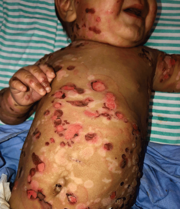
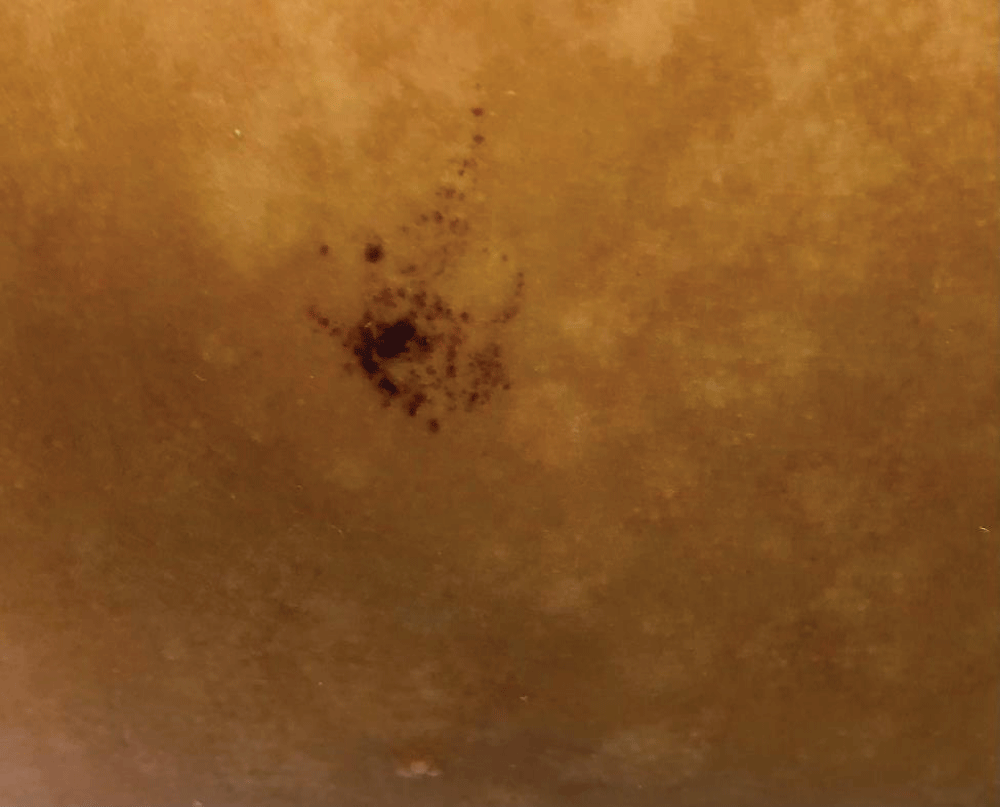
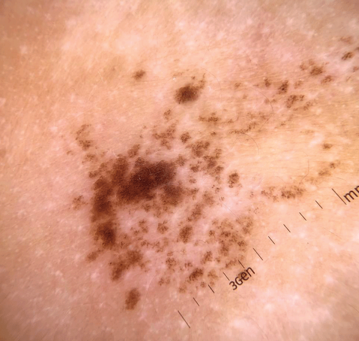
Epidermolysis Bullosa Naevi (EBN) is a subset of melanocytic nevi with atypical features arising at sites of blistering in patients with congenital EB. It may be clinically misdiagnosed as melanoma and may represent a challenge for the dermatologist. Bullous Pemphigoid (BP) consists of an autoimmune condition presenting with subepidermal blisters, usually affecting the elderly and rarely observed in children The case is reported of an infant who presented with pruritic erythematous bullous lesions, initially appearing over the trunk and legs with progression to arms and face. Clinical and immunopathological features were consistent with the diagnosis of infant BP. In the course of the disease, he developed a pigmented heterogeneous macule with irregular contour and satellite-dotted lesions, located on the right flank. Dermoscopy revealed a regular pigmented network distributed in an agminated manner interspersed with areas of healthy skin. Due to its similarity to EBN, an expectant approach was carried out. The lesion regressed during the 24-month follow-up. To our knowledge, there is only one literature case report of a child who presented with EBN-like in a previous BP lesion. Our case reinforces the presence of atypical melanocytic nevi in bullous diseases. Knowing this type of lesion clinically and dermatoscopically in patients with bullous dermatoses may prevent unnecessary surgical procedures in children.


